

Relationship between grades of macular perfusion and foveal thickness in branch retinal vein occlusion
- PMID: 23308037
- PMCID: PMC3538501
- DOI: 10.2147/OPTH.S37185
Relationship between grades of macular perfusion and foveal thickness in branch retinal vein occlusion
Abstract
Background: To study the relationship between retinal perfusion around the macula and the foveal thickness in branch retinal vein occlusion (BRVO).
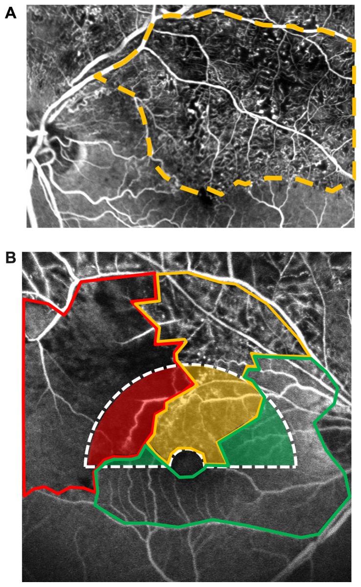
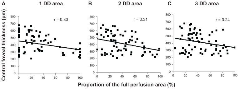
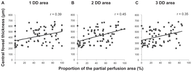
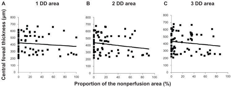
Methods: Seventy-four eyes of 74 consecutive patients with BRVO were enrolled. We developed a new grading system to evaluate the status of retinal perfusion around the macula in three grades: full perfusion area (FPA), partial perfusion area (PPA), and nonperfusion area (NPA), using fluorescein angiography. The correlation was assessed between these grades and the central foveal thickness (CFT) measured by optical coherence tomography. We also determined the area with the closest correlation between the perfusion status and the foveal thickness by comparing the correlation coefficient in areas of 1-, 2-, and 3-disc diameter (DD) horizontal hemicircles centered at the fovea. The correlation was determined between the extent of each perfusion grade and CFT.
Results: We found a significant negative correlation between the CFT and the FPA (r = 0.31, P = 0.006) and a significant positive correlation between the CFT and the PPA (r = 0.45, P < 0.001) in the three areas. The most significant correlations were found in the 2-DD area. Interestingly, the NPA has not correlated with the foveal thickness in any areas.
Conclusion: The areas of partial but not complete capillary loss seem to be responsible for the macular edema associated with BRVO. Treatments targeting leakage from the dilated capillaries in the PPA should be investigated.
Keywords: branch retinal vein occlusion; foveal thickness; macular edema; macular perfusion.
Figures
References
-
- Branch Vein Occlusion Study Group. Argon laser scatter photocoagulation for prevention of neovascularization and vitreous hemorrhage in branch vein occlusion: a randomized clinical trial. Arch Ophthalmol. 1986;104(1):34–41. - PubMed
-
- Rogers SL, McIntosh RL, Lim L, et al. Natural history of branch retinal vein occlusion: an evidence-based systematic review. Ophthalmology. 2010;117(6):1094–1101. - PubMed
-
- Branch Vein Occlusion Study Group. Argon laser photocoagulation for macular edema in branch vein occlusion. Am J Ophthalmol. 1984;98(3):271–282. - PubMed
-
- Michels RG, Gass JD. The natural course of retinal branch vein obstruction. Trans Am Acad Ophthalmol Otolaryngol. 1974;78(2):166–177. - PubMed
LinkOut - more resources
Full Text Sources
