. 2012 Dec;14(12):1164-77.
doi: 10.1593/neo.121088.

E-cadherin controls bronchiolar progenitor cells and onset of preneoplastic lesions in mice

Affiliations

E-cadherin controls bronchiolar progenitor cells and onset of preneoplastic lesions in mice

Fatih Ceteci et al. Neoplasia. 2012 Dec.

Abstract

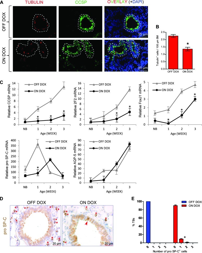
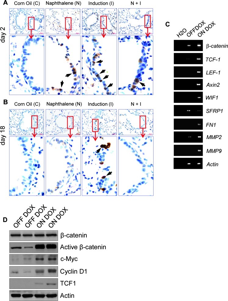
Although progenitor cells of the conducting airway have been spatially localized and some insights have been gained regarding their molecular phenotype, relatively little is known about the mechanisms regulating their maintenance, activation, and differentiation. This study investigates the potential roles of E-cadherin in mouse Clara cells, as these cells were shown to represent the progenitor/stem cells of the conducting airways and have been implicated as the cell of origin of human non-small cell lung cancer. Postnatal inactivation of E-cadherin affected Clara cell differentiation and compromised airway regeneration under injury conditions. In steady-state adult lung, overexpression of the dominant negative E-cadherin led to an expansion of the bronchiolar stem cells and decreased differentiation concomitant with canonical Wnt signaling activation. Expansion of the bronchiolar stem cell pool was associated with an incessant proliferation of neuroepithelial body.associated Clara cells that ultimately gave rise to bronchiolar hyperplasia. Despite progressive hyperplasia, only a minority of the mice developed pulmonary solid tumors, suggesting that the loss of E-cadherin function leads to tumor formation when additional mutations are sustained. The present study reveals that E-cadherin plays a critical role in the regulation of proliferation and homeostasis of the epithelial cells lining the conducting airways.


Figures

Figure 1
Figure 1
Conditional inactivation of E-cadherin in steady-state lungs alters Clara cell differentiation. (A) Construct and breeding strategy used to inactivate E-cadherin in Clara cells in the conducting airway. m, myc tag. (B) Immunoblot analysis shows transgene expression only in the presence of DOX. Actin was used as a control. (C) Immunohistochemical staining of lung sections for myc tag. Five-week-old DTR mice were either untreated (OFF DOX) or DOX-treated (ON DOX) for 1 week before analysis. Transgene expression was detected both in conducting airways (red circles) and in the alveoli (green circles). Arrows in C indicate regions shown in insets. Hematoxylin (blue) was used as a counterstain. Scgb1a1/myc tag (D) and Scgb1a1/E-cadherin (E) dual immunofluorescence stainings of lung sections from control and induced adult mice. Myc tag.expressing Clara cells are shown with orange arrows. White arrows in D show Scgb1a1-negative but myc tag. positive cells. Arrows in E indicate regions shown in insets. Nuclei were counterstained with 4′,6-diamidino-2-phenylindole (DAPI, blue).
Figure 2
Figure 2
Disruption of E-cadherin in Scgb1a1-expressing cells impairs Clara cell lineage differentiation and induces ectopic expression of pro SpC in conducting airways. (A) Representative dual immunofluorescence staining of lung sections for tubulin (red) and Scgb1a1 (green). Individual bronchioles are encircled by dashed lines. Nuclei were counterstained with DAPI (blue). (B) Morphometric analysis of ciliated cells. Results are presented as means ± SEM for each group. *P < .05. BM, basement membrane. (C) Gene expression as a function of age. Total lung RNA from control and induced mice was assayed for expression of Scgb1a1, cytochrome P450-2F2, FoxJ1, SpC, and AQP-5 by quantitative RT-PCR, and average ddCT values are presented ± SEM. NB, new born. (D) Immunohistochemistry shows ectopic expression of the alveolar type II cell marker pro Sftpc (red arrow) in bronchiolar epithelium of an induced mouse. Type II pneumocytes (red asterisks) served as an internal control for pro Sftpc staining (brown). Hematoxylin (blue) was used as a counterstain. (E) The percentage of terminal bronchioles containing zero, one, and two cells with ectopic pro Sftpc expression is shown. At least 220 bronchioles were counted from six DTR mice for each group. Results are presented as means ± SEM for each group. *P < .05.
Figure 3
Figure 3
E-cadherin is necessary for the repair of conducting airway epithelium after injury. Five-week-old mice were treated as indicated. Mice were recovered following 2 or 18 days from naphthalene or corn oil exposures. (A) Immunohistochemical staining of lung sections for Scgb1a1. Images are representative of n = 4 separate mice per treatment group. Br, bronchiole. (B) Double immunofluorescence staining of a lung section for Scgb1a1 (red) and Ki67 (green) shows actively proliferating naphthalene-resistant Clara cells in bronchiolar epithelium of a medium airway (dotted line) 2 days after naphthalene treatment. Nuclei were counterstained with DAPI (blue). (C) Immunohistochemical staining of lungs sections for CYP2F2. Red arrows indicate naphthalene-resistant Clara cells. Black arrows and green brackets indicate Scgb1a1-negative regions. Insets represent higher magnification of the areas shown in boxed areas. Blue circles indicate increased cellular densities. Hematoxylin (blue) was used as a counterstain. (D and E) Morphometric quantification of the epithelial damage at day 2 and day 18 after naphthalene-mediated injury. The impact of treatment on epithelial repair was determined through analysis of Scgb1a1 (E) or CYP2F2 (D) repopulation. Results are presented as means ± SEM for each group (n = 4). *P < .05. BM, basement membrane.
Figure 4
Figure 4
Analysis of cell proliferation within NEB regions under airway repair and steady-state conditions after E-cadherin inactivation. (A) Triple immunofluorescence staining of lung sections for indicated markers. Naphthalene-resistant Clara cells (ClaraV) were identified with their close association with CGRP-expressing neuroendocrine cells in NEBs. Asterisks show proliferating Clara cells that are NEB-independent. Nuclei were counterstained with DAPI (blue). Quantitation of cell proliferation for PNECs (B) and for ClaraV cells (C) for indicated treatment groups. Results are presented as average ± SEM for each group (n = 4). *P < .05.
Figure 5
Figure 5
Clara cell.specific loss of E-cadherin in adult lungs leads to ectopic BASC-like progenitor cell localization. (A) Representative double immunofluorescence staining of lung sections for pro Sftpc (green) and Scgb1a1 (red) shows ectopic BASC-like progenitor cell localizations (arrows) in an induced mouse compared to control. Yellow arrow indicates the region shown in the inset. Nuclei were counterstained with DAPI (blue). (B) Gating strategy for FACS sorting. Cells were first gated on the CD31/CD45neg and CD34pos population and then subsequently gated on the Sca1pos population. (C) Quantitation of flow cytometry for BASC-like progenitor cells between the uninduced (OFF DOX) and 1-month DOX-induced (ON DOX) DTR mice (n = 3 mice for each group). Results are presented as means ± SEM for each group. ns, statistically not significant.
Figure 6
Figure 6
Activation of canonical Wnt signaling upon airway injury or as a consequence of dn E-cadherin expression in conducting airway epithelium. (A and B) Immunohistochemistry of lung sections for β-catenin. Duration and type of treatment as indicated. Highmagnification images represent regions shown in insets. Arrows show nuclear staining of β-catenin (brown). Hematoxylin (blue) was used as a counterstain. (C) Semiquantitative RT-PCR analysis of Wnt/β-catenin.regulated genes for control (OFF DOX) and induced (ON DOX) DTR mice. Actin was used as an internal control. (D) Immunoblot analysis of total lung lysates of control and induced DTR mice with the indicated markers.
Figure 7
Figure 7
Loss of E-cadherin expression in conducting airway epithelium leads to bronchiolar hyperplasia. (A) Histologic analysis through H&E staining of lungs from control and 2-week DOX-treated DTR mice showing the presence of epithelial hyperplasia (arrow). (B) Quantitation of bronchiolar hyperplasia between the mice groups: wt, wild type (n = 13), (C) Scgb1a1-rtTA (n = 27), (D) Tet-O dn E-cadherin (n = 23), DTR OFF DOX (n = 39), DTR ON DOX (n = 40); *P < .05. (C) Double immunofluorescence staining of the same lung sections from A for indicated markers. Asterisk shows a PGP 9.5-positive NEB region. Marked region shows an expansion of an NEB region consisting of PGP 9.5-and Scgb1a1-positive cells after induction. Nuclei were counterstained with DAPI (blue). (D) Immunohistochemical staining of lung sections for Scgb1a1. Images are representative of n = 6 mice in each group. Hematoxylin (blue) was used as a counterstain.
Figure 8
Figure 8
Analysis of incidence and histopathology of pulmonary tumors from induced transgenic mice. (A) Lung sections from tumor-bearing mice were either H&E stained or immunostained for Ki67 (brown). Arrows indicate tumor foci. (B) Immunohistochemistry of tumor-bearing lung sections for lung differentiation markers as indicated. Arrows show rare Scgb1a1-positive tumor cells that were never detected in spontaneous lung tumor but in their surrounding bronchioles. (C and D) Staining of lung tumor sections with the indicated markers. Arrows in C show Alcian Blue.positive vessel and those in D show vimentin-positive vessel. Two representative pictures were shown for each staining.

Similar articles

Cited by

References

    1. Maeda Y, Dave V, Whitsett JA. Transcriptional control of lung morphogenesis. Physiol Rev. 2007;87:219–244. - PubMed
    1. Blenkinsopp WK. Proliferation of respiratory tract epithelium in the rat. Exp Cell Res. 1967;46:144–154. - PubMed
    1. Evans MJ, Cabral LC, Stephens RJ, Freeman G. Acute kinetic response and renewal of the alveolar epithelium following injury by nitrogen dioxide. Chest. 1974;65(suppl):62S–65S. - PubMed
    1. Evans MJ, Cabral LJ, Stephens RJ, Freeman G. Transformation of alveolar type 2 cells to type 1 cells following exposure to NO2. Exp Mol Pathol. 1975;22:142–150. - PubMed
    1. Hong KU, Reynolds SD, Watkins S, Fuchs E, Stripp BR. Basal cells are a multipotent progenitor capable of renewing the bronchial epithelium. Am J Pathol. 2004;164:577–588. - PMC - PubMed

Supplementary References

    1. Dahl U, Sjodin A, Semb H. Cadherins regulate aggregation of pancreatic β-cells in vivo. Development. 1996;122:2895–2902. - PubMed
    1. Liu C, Glasser SW, Wan H, Whitsett JA. GATA-6 and thyroid transcription factor-1 directly interact and regulate surfactant protein-C gene expression. J Biol Chem. 2002;277:4519–4525. - PubMed
    1. Van Winkle LS, Gunderson AD, Shimizu JA, Baker GL, Brown CD. Gender differences in naphthalene metabolism and naphthalene-induced acute lung injury. Am J Physiol Lung Cell Mol Physiol. 2002;282:L1122–L1134. - PubMed
    1. Wu CA, Peluso JJ, Shanley JD, Puddington L, Thrall RS. Murine cytomegalovirus influences Foxj1 expression, ciliogenesis, and mucus plugging in mice with allergic airway disease. Am J Pathol. 2008;172:714–724. - PMC - PubMed
    1. Rapp UR, Korn C, Ceteci F, Karreman C, Luetkenhaus K, Serafin V, Zanucco E, Castro I, Potapenko T. MYC is a metastasis gene for non-small-cell lung cancer. PLoS One. 2009;4:e6029. - PMC - PubMed

Publication types

MeSH terms

LinkOut - more resources