. 2012 Dec;8(12):e1003097.
doi: 10.1371/journal.ppat.1003097. Epub 2012 Dec 20.

4'-Phosphopantetheinyl transferase PptT, a new drug target required for Mycobacterium tuberculosis growth and persistence in vivo

Affiliations

4'-Phosphopantetheinyl transferase PptT, a new drug target required for Mycobacterium tuberculosis growth and persistence in vivo

Cécile Leblanc et al. PLoS Pathog. 2012 Dec.

Abstract

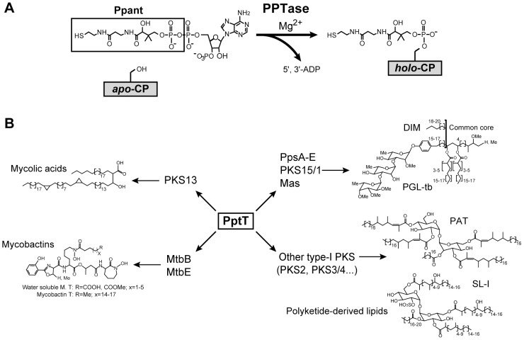
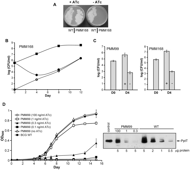
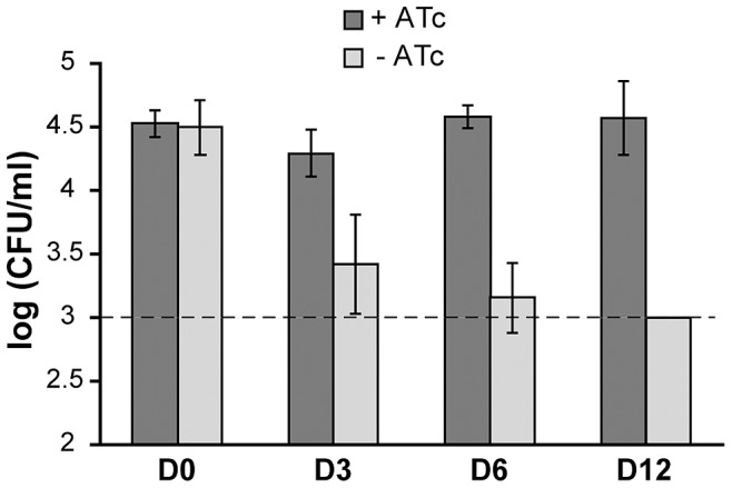
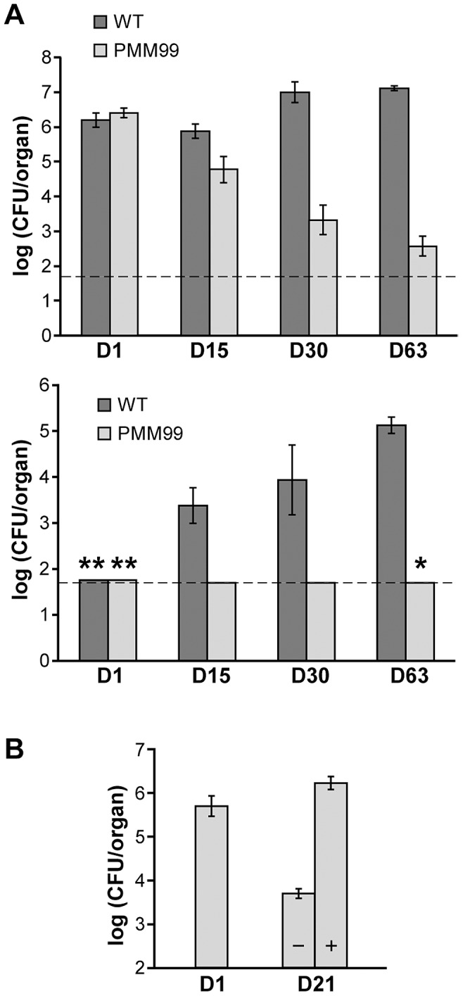
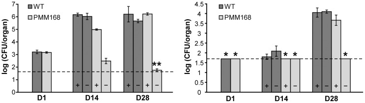
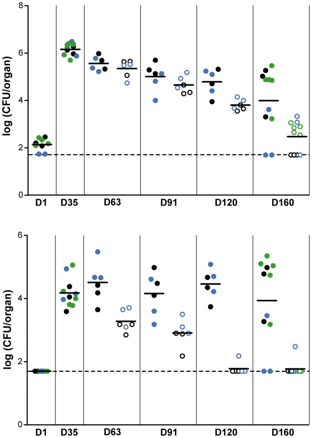
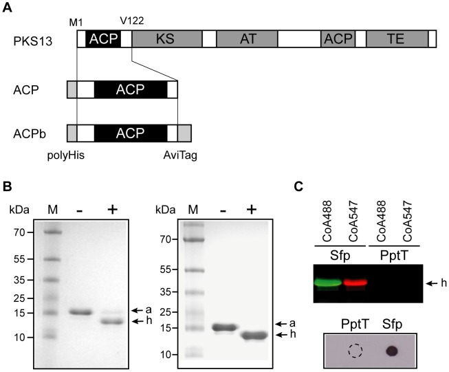
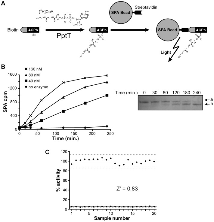
The cell envelope of Mycobacterium tuberculosis, the causative agent of tuberculosis in humans, contains lipids with unusual structures. These lipids play a key role in both virulence and resistance to the various hostile environments encountered by the bacteria during infection. They are synthesized by complex enzymatic systems, including type-I polyketide synthases and type-I and -II fatty acid synthases, which require a post-translational modification to become active. This modification consists of the covalent attachment of the 4'-phosphopantetheine moiety of Coenzyme A catalyzed by phosphopantetheinyl transferases (PPTases). PptT, one of the two PPTases produced by mycobacteria, is involved in post-translational modification of various type-I polyketide synthases required for the formation of both mycolic acids and lipid virulence factors in mycobacteria. Here we identify PptT as a new target for anti-tuberculosis drugs; we address all the critical issues of target validation to demonstrate that PptT can be used to search for new drugs. We confirm that PptT is essential for the growth of M. bovis BCG in vitro and show that it is required for persistence of M. bovis BCG in both infected macrophages and immunodeficient mice. We generated a conditional expression mutant of M. tuberculosis, in which the expression of the pptT gene is tightly regulated by tetracycline derivatives. We used this construct to demonstrate that PptT is required for the replication and survival of the tubercle bacillus during the acute and chronic phases of infection in mice. Finally, we developed a robust and miniaturized assay based on scintillation proximity assay technology to search for inhibitors of PPTases, and especially of PptT, by high-throughput screening. Our various findings indicate that PptT meets the key criteria for being a therapeutic target for the treatment of mycobacterial infections.


Conflict of interest statement

The authors have declared that no competing interests exist.

Figures

Figure 1
Figure 1. Role of PptT in M. tuberculosis.
A. Enzymatic reaction catalyzed by PPTases. CP: carrier protein. B. Schematic diagram of the role of PptT in the biosynthesis pathways for mycolic acids, polyketide-derived lipids and siderophores in M. tuberculosis. DIM: phthiocerol dimycocerosates, PGL: phenolglycolipids, PAT: polyacyltrehaloses, SL: sulfolipids.
Figure 2
Figure 2. Effect of PptT depletion on the growth of M. bovis BCG and of M. tuberculosis in vitro.
A. M. tuberculosis H37Rv wild-type (WT) and the PMM168 mutant strain were grown in 7H9 containing ADC (with Km, Hyg and ATc for PMM168) at 37°C and streaked onto 7H11 plates supplemented with OADC with or without ATc (300 ng/ml). Plates were incubated for 20 days at 37°C. B. The M. tuberculosis PMM168 mutant was grown in 7H9 containing or not containing ATc at 37°C. Numbers of CFU in cultures with ATc (squares) were determined by plating dilutions of theses cultures onto 7H11 plates supplemented with ATc on days 0, 4, 8 and 12. CFU counts in cultures lacking ATc were determined by plating dilutions on 7H11 plates supplemented with ATc (closed circles) or without ATc (open circles) to estimate the number of ATc-independent CFU. C. Bactericidal effect of PptT depletion. The M. bovis BCG PMM99 (left panel) and M. tuberculosis PMM168 (right panel) mutants were grown in 7H9 containing (+) or not containing (−) ATc at 37°C. Numbers of CFU in cultures were determined by plating dilutions of theses cultures onto 7H11 plates supplemented with ATc on days 0 (D0) and 4 (D4). Values are means ± standard deviations (error bars) of CFU counts for three independent experiments. D. PMM99 was grown in media supplemented with Tween-80 with (100, 1, 0.3, 0.1 ng/ml) and without ATc and bacterial growth was monitored by measuring the optical density at 600 nm (OD600) (left panel). M. bovis BCG wild-type strain was grown in 7H9 supplemented with Tween-80. Data are representative of two independent experiments. Western blot visualization of PptT in crude cell lysates of PMM99 (5 µg/lane) cultivated for 6 days in a medium with ATc (100, 1, 0.3 ng/ml) and in a crude cell lysate of M. bovis BCG wild-type strain (5, 2, 1, 0.5 µg/lane) (right panel). The control lane was loaded with 100 ng of recombinant PptT fused to a poly-histidine tag produced in E. coli.
Figure 3
Figure 3. Effect of PptT depletion on survival of PMM99 in murine macrophages.
Bone marrow-derived macrophages were infected with strain PMM99, cultivated in the presence or absence of ATc (500 ng/ml) and lysed after 0, 3, 6 or 12 days. Viable bacteria were counted by plating dilutions of the lysates on selective media. The data reported are from one experiment performed in triplicate and representative of three independent experiments. The dashed line corresponds to the detection limit (3 log, see materials and methods).
Figure 4
Figure 4. SCID mice infections with the PMM99 mutant.
A. Competition between strain PMM99 and wild-type M. bovis BCG in infected SCID mice. SCID mice were infected with a mixture of wild-type M. bovis BCG and PMM99. Numbers of CFU of each strain recovered from the lungs (upper panel) and the spleen (lower panel) of SCID mice 1 day (D1), 15 days (D15), 30 days (D30) and 63 days (D63) after infection were determined by plating dilutions of homogenized tissues on selective media. Values are means ± standard deviations (error bars) of CFU counts for 4 (day 63) or 5 (days 1, 15, 30) infected mice. The dashed lines correspond to the detection limit (1.7 log, see materials and methods). Asterisks indicate that CFU counts in all (*) or some (**) infected mice were below the detection limit (50 CFU/organ): the number of CFU scored in such cases is 50 CFU per organ. The average number of CFU recovered from the organ is therefore overestimated. B. Effect of doxycycline treatment on the survival of PMM99 in SCID mice. SCID mice were infected with strain PMM99 and administered or not administered 0.1 mg/ml doxycycline. Numbers of CFU recovered from the lungs of infected mice on day 1 (D1) and from the lungs of untreated (−) and treated (+) mice on day 21 (D21) were determined by plating dilutions of homogenized tissues on 7H11 media. Values are means ± standard deviations (error bars) of CFU counts for three infected mice.
Figure 5
Figure 5. Effect of PptT depletion on the multiplication of M. tuberculosis H37Rv in BALB/c mice.
Mice were infected either with the wild-type M. tuberculosis strain or with strain PMM168 and either received or did not receive doxycycline treatment (0.1 mg/ml) from one day post-infection. Numbers of CFU of M. tuberculosis wild-type (dark gray bars) or of PMM168 (light gray bars) in lungs (left panel) and spleen (right panel) of treated (+) and untreated (−) mice were determined on days 1 (D1), 14 (D14) and 28 (D28) by plating dilutions of homogenized tissues on selective media. Values are means ± standard deviations (error bars) of CFU counts for three or four infected mice (see materials and methods). The dashed line corresponds to the detection limit (1.7 log). Asterisks indicate the counts in all (*) or some (**) infected mice were below the detection limit (50 CFU/organ). In such cases, the number of CFU scored was 50 CFU per organ; consequently mean number of CFU per organ is an overestimation.
Figure 6
Figure 6. Effect of PptT depletion on the persistence of M. tuberculosis H37Rv in BALB/c mice.
Mice were infected with strain PMM168 and treated with 0.1 mg/ml doxycycline from 1 day post-infection. Thirty-five days post-infection mice were split in two groups: group 1 continued to receive doxycycline and doxycycline treatment was withdrawn from group 2. Numbers of CFU present in lungs (upper panel) and spleen (lower panel) of treated (closed circles) and of untreated (open circles) mice were determined 1 (D1), 35 (D35), 63 (D63), 91 (D91), 120 (D120) and 160 (D160) days post-infection (experiments 1 and 2, black and blue circles) or 1, 35 and 160 days post-infection (experiment 3, green circles). Each circle represents CFU obtained from one mouse. The dashed line corresponds to the detection limit. When counts in infected mice were below the detection limit, the number of CFU scored was 50 CFU per organ (1.7 log).
Figure 7
Figure 7. In vitro activity of PptT.
A. Diagrammatic representation of the domain organization of PKS13 and of ACP and ACPb domains. ACP: Acyl Carrier protein, KS: ketosynthase, AT: acyltransferase, TE: thioesterase. B. ACP activation with CoA and acetyl-CoA. The apo-ACP module was incubated with (+) or without (−) PptT in the presence of either CoA (left panel) or acetyl-CoA (right panel). apo- (a) and holo-ACP (h) forms were separated on urea polyacrylamide gels and stained with Coomassie blue (see materials and methods). M: PageRuler prestained protein ladder plus (Fermentas). C. ACP activation with CoA analogs. PptT or Sfp were incubated with the apo-ACP domain in the presence of either fluorescent CoA analogs (CoA488 or CoA547) or CoA-biotin. Fluorescent holo-ACP forms (h) were resolved by SDS-PAGE and visualized by fluorescence scanning using a Typhoon scanner (GE Healthcare) (upper panel). Biotin-labeled ACP was detected by spotting 5 µl of the reaction mix onto the nitrocellulose membrane and incubation with streptavidin peroxidase followed by enhanced chemiluminescence detection (lower panel). The dashed-line circle shows the drop zone for the PptT reaction.
Figure 8
Figure 8. PptT SPA assay for high throughput screening.
A. Principle of the PptT SPA assay. B. Effect of enzyme concentration on the SPA assay signal. Biotinylated apo-ACPb (4 µM) and radiolabeled [3H]CoA (2 µM) were incubated in the presence of 0, 40, 80, or 160 nM of PptT in standard conditions (see materials and methods). Reactions were stopped after various times by addition of stop buffer and 250 µg of SPA beads resuspended in 60 µl of water. Signals were detected by scintillation counting using a TopCount (Perkin Elmer). A parallel kinetic experiment was carried out with 80 nM of enzyme: the reaction was stopped at various times and loaded onto a urea polyacrylamide gel to separate apo- (a) and holo-ACP (h) forms (right panel). The gel was stained with Coomassie blue. C. Determination of the Z′ factor. SPA assays were carried out in standard conditions (4 µM of ACPb, 2 µM of [3H]CoA, 2 µM of CoA) in the presence of 80 nM of enzyme (n = 20, black squares) or without enzyme (n = 20, black circles) at 30°C for 1.5 hour in a 96-well plate. Reactions were stopped by addition of stop buffer and SPA beads, and scintillation signals were detected using a TopCount. The Z′ factor was calculated as indicated in materials and methods (μc+ = 100, μc− = 5.53, σc+ = 4.74, σc− = 0.42). The dotted lines correspond to three standard deviations from the mean of the positive and negative controls. Data are expressed relative to the mean value for positive controls.

Similar articles

Cited by

References

    1. Almeida Da Silva PE, Palomino JC (2011) Molecular basis and mechanisms of drug resistance in Mycobacterium tuberculosis: classical and new drugs. J Antimicrob Chemoth 66: 1417–1430. - PubMed
    1. Russell DG (2011) Mycobacterium tuberculosis and the intimate discourse of a chronic infection. Immunol Rev 240: 252–268. - PMC - PubMed
    1. Pieters J (2008) Mycobacterium tuberculosis and the macrophage: maintaining a balance. Cell Host Microbe 3: 399–407. - PubMed
    1. Stewart GR, Robertson BD, Young DB (2003) Tuberculosis: a problem with persistence. Nat Rev Microbiol 1: 97–105. - PubMed
    1. Betts JC, Lukey PT, Robb LC, McAdam RA, Duncan K (2002) Evaluation of a nutrient starvation model of Mycobacterium tuberculosis persistence by gene and protein expression profiling. Mol Microbiol 43: 717–731. - PubMed

MeSH terms

Substances