. 2013;8(1):e52323.
doi: 10.1371/journal.pone.0052323. Epub 2013 Jan 7.

A unified model of the GABA(A) receptor comprising agonist and benzodiazepine binding sites

Affiliations

A unified model of the GABA(A) receptor comprising agonist and benzodiazepine binding sites

Rikke Bergmann et al. PLoS One. 2013.

Abstract

We present a full-length α(1)β(2)γ(2) GABA receptor model optimized for agonists and benzodiazepine (BZD) allosteric modulators. We propose binding hypotheses for the agonists GABA, muscimol and THIP and for the allosteric modulator diazepam (DZP). The receptor model is primarily based on the glutamate-gated chloride channel (GluCl) from C. elegans and includes additional structural information from the prokaryotic ligand-gated ion channel ELIC in a few regions. Available mutational data of the binding sites are well explained by the model and the proposed ligand binding poses. We suggest a GABA binding mode similar to the binding mode of glutamate in the GluCl X-ray structure. Key interactions are predicted with residues α(1)R66, β(2)T202, α(1)T129, β(2)E155, β(2)Y205 and the backbone of β(2)S156. Muscimol is predicted to bind similarly, however, with minor differences rationalized with quantum mechanical energy calculations. Muscimol key interactions are predicted to be α(1)R66, β(2)T202, α(1)T129, β(2)E155, β(2)Y205 and β(2)F200. Furthermore, we argue that a water molecule could mediate further interactions between muscimol and the backbone of β(2)S156 and β(2)Y157. DZP is predicted to bind with interactions comparable to those of the agonists in the orthosteric site. The carbonyl group of DZP is predicted to interact with two threonines α(1)T206 and γ(2)T142, similar to the acidic moiety of GABA. The chlorine atom of DZP is placed near the important α(1)H101 and the N-methyl group near α(1)Y159, α(1)T206, and α(1)Y209. We present a binding mode of DZP in which the pending phenyl moiety of DZP is buried in the binding pocket and thus shielded from solvent exposure. Our full length GABA(A) receptor is made available as Model S1.


Conflict of interest statement

Competing Interests: TS is employed at Novo Nordisk A/S. Post Doc RB was funded by The Velux Foundation and Reseach Assistant KK was funded by Neurosearch A/S. This does not alter the authors' adherence to all the PLoS ONE policies on sharing data and materials.

Figures

Figure 1
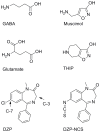
Figure 1. Some classical GABAA receptor ligands.
GABA is the endogenous GABAR agonist, muscimol a classical high-affinity agonist and THIP a muscimol analogue. Although not a GABAR ligand, glutamate is included to illustrate the resemblance to GABA. Diazepam (DZP) belongs to the benzodiazepine class of compounds, which are allosteric GABAA modulators. The DZP-NCS analogue attaches covalently to GABAARs and is included for validation purposes.
Figure 2
Figure 2. Illustration of the GABAAR structural composition.
A) Top view showing the pentameric assembly of α1, β2 and γ2 subunits and the location of binding sites for GABA and BZDs; B) Side view illustrating the extracellular domain (ECD) where agonists and benzodiazepines bind and the transmembrane domain (TMD); C) Zooming in on a GABA binding site at the subunit interface between β2 and α1 subunits, loop regions A–F mentioned in the text are shown (A: yellow, B: orange, C: red, D: purple, E: blue and F: pink).
Figure 3
Figure 3. Alignment of protein sequences from GluCl, GLIC, ELIC and the human α1, β2, and γ2 GABAAR subunits.
The GluCl sequence was used as template for homology modeling throughout the GABAAR subunits, and ELIC was included as a template in the regions marked with blue boxes. The secondary structure deduced from the X-ray structure of GluCl is shown above the alignment (red shapes denote α-helices, and green arrows denote β-strands), whereas historically assigned loop regions are indicated below the alignment. As in the GluCl structure the M3–M4 intracellular loop of GABAAR sequences was replaced by an AGT tri-peptide linker. Residues comprising the binding sites (within 8 Å of Glu in the GluCl structure and pointing towards the binding site) are colored pink (Glu and GABA binding site) and cyan (BZD binding site). Binding site residues conserved with respect to the templates are indicated as follows: + conserved in both GABA and BZD binding sites; * conserved in the GABA binding sites; . conserved in the BZD binding site. For details on calculation of binding site sequence identities see Figure S1.
Figure 5
Figure 5. Agonist binding modes determined by induced fit docking.
A) GABA (green), B) THIP (pink) and C) muscimol (cyan) are shown in the orthosteric binding site at the interface between the β2 subunit (teal) and the α1 subunit (smudge).
Figure 4
Figure 4. Conserved template residues.
The figure shows residues that are conserved or homologous to GABAAR binding site residues from the GluCl X-ray structure (PBD ID 3RIF) as grey sticks and the bacterial Cys-Loop receptor homolog, ELIC (PDB ID 2VL0) as purple sticks. Glutamate as co-crystallized with GluCl is shown in yellow, where the structure corresponding to GABA is shown as sticks and the α-carboxylic acid removed prior to homology modeling is shown as lines. A) GABA binding site and B) BZD binding site.
Figure 6
Figure 6. Conformational energy profile for dihedral drive of the amino-methyl side chain of muscimol.
B3LYP/6-31G** energies.
Figure 7
Figure 7. Region suited for a tightly bound water molecule identified in agonist site.
A GRID calculation at the agonist binding site, using the water probe, identified two regions of strong binding interaction energy (−11 kcal/mol). One region is overlapping with the acidic moiety of agonists and the other region is situated next to the backbone of β2S156 and β2Y157 (grey mesh). The calculation was performed in absence of agonist in the binding site. In the picture, the site has been optimized for muscimol as described in the methods section. A) When a water molecule is placed between muscimol and the B-loop backbone, perfect hydrogen bonding distances are obtained, resulting in optimal interactions between the high affinity ligand muscimol and the GABA receptor. B) When also GABA is included in the site, it is obvious that the water molecule would make up for the backbone interaction that GABA is predicted to make.
Figure 8
Figure 8. DZP binding mode.
A) The assumed biologically active binding mode of DZP (gray) at the interface between the α1 (smudge) and γ2 (firebrick) subunits. In this conformation the C-3 points upwards and the pending phenyl substituent is directed inwards. B) Covalently attached DZP-NCS (cyan) overlaid with DZP (gray). Only moderate differences between the docked and the covalently attached ligands exist.

References

    1. Johnston GA (2005) GABA(A) receptor channel pharmacology. Curr Pharm Des 11: 1867–1885. - PubMed
    1. Johnston GA, Curtis DR, De Groat WC, Duggan AW (1968) Central actions of ibotenic acid and muscimol. Biochem Pharmacol 17: 2488–2489. - PubMed
    1. Ebert B, Frolund B, Diemer NH, Krogsgaard-Larsen P (1999) Equilibrium binding characteristics of [3H]thiomuscimol. Neurochem Int 34: 427–434. - PubMed
    1. Krogsgaard-Larsen P, Johnston GA, Lodge D, Curtis DR (1977) A new class of GABA agonist. Nature 268: 53–55. - PubMed
    1. Frolund B, Kristiansen U, Brehm L, Hansen AB, Krogsgaard-Larsen P, et al. (1995) Partial GABAA receptor agonists. Synthesis and in vitro pharmacology of a series of nonannulated analogs of 4,5,6,7-tetrahydroisoxazolo[5,4-c]pyridin-3-ol. J Med Chem 38: 3287–3296. - PubMed

Publication types

LinkOut - more resources