

Saffold virus type 3 (SAFV-3) persists in HeLa cells
- PMID: 23308162
- PMCID: PMC3537732
- DOI: 10.1371/journal.pone.0053194
Saffold virus type 3 (SAFV-3) persists in HeLa cells
Erratum in
- PLoS One. 2013;8(9):doi/10.1371/annotation/33ee31ba-3163-4859-ac37-7300ea78537e
Abstract
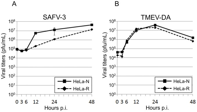
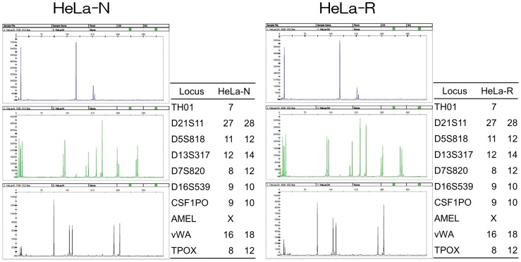
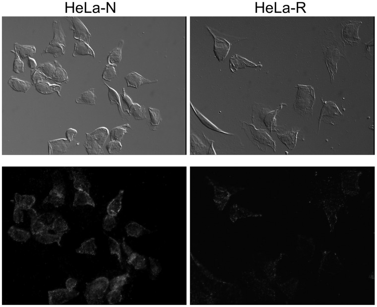
Saffold virus (SAFV) was identified as a human cardiovirus in 2007. Although several epidemiological studies have been reported, they have failed to provide a clear picture of the relationship between SAFV and human diseases. SAFV genotype 3 has been isolated from the cerebrospinal fluid specimen of patient with aseptic meningitis. This finding is of interest since Theiler's murine encephalomyelitis virus (TMEV), which is the closely related virus, is known to cause a multiple sclerosis-like syndrome in mice. TMEV persistently infects in mouse macrophage cells in vivo and in vitro, and the viral persistence is essential in TMEV-induced demyelinating disease. The precise mechanism(s) of SAFV infection still remain unclear. In order to clarify the SAFV pathogenicity, in the present study, we studied the possibilities of the in vitro persistent infection of SAFV. The two distinct phenotypes of HeLa cells, HeLa-N and HeLa-R, were identified. In these cells, the type of SAFV-3 infection was clearly different. HeLa-N cells were lyticly infected with SAFV-3 and the host suitable for the efficient growth. On the other hand, HeLa-R cells were persistently infected with SAFV-3. In addition, the SAFV persistence in HeLa-R cells is independent of type I IFN response of host cells although the TMEV persistence in mouse macrophage cells depends on the response. Furthermore, it was suggested that SAFV persistence may be influenced by the expression of receptor(s) for SAFV infection on the host cells. The present findings on SAFV persistence will provide the important information to encourage the research of SAFV pathogenicity.
Conflict of interest statement
Figures
Similar articles
-
Saffold virus, a novel human Cardiovirus with unknown pathogenicity.J Virol. 2012 Feb;86(3):1292-6. doi: 10.1128/JVI.06087-11. Epub 2011 Nov 23. J Virol. 2012. PMID: 22114344 Free PMC article. Review.
-
Saffold virus, a human Theiler's-like cardiovirus, is ubiquitous and causes infection early in life.PLoS Pathog. 2009 May;5(5):e1000416. doi: 10.1371/journal.ppat.1000416. Epub 2009 May 1. PLoS Pathog. 2009. PMID: 19412527 Free PMC article.
-
The Pathogenesis of Saffold Virus in AG129 Mice and the Effects of Its Truncated L Protein in the Central Nervous System.Viruses. 2016 Feb 18;8(2):24. doi: 10.3390/v8020024. Viruses. 2016. PMID: 26901216 Free PMC article.
-
Cultivation and serological characterization of a human Theiler's-like cardiovirus associated with diarrheal disease.J Virol. 2010 May;84(9):4407-14. doi: 10.1128/JVI.02536-09. Epub 2010 Feb 17. J Virol. 2010. PMID: 20164225 Free PMC article.
-
Saffold virus, an emerging human cardiovirus.Rev Med Virol. 2017 Jan;27(1):e1908. doi: 10.1002/rmv.1908. Epub 2016 Oct 10. Rev Med Virol. 2017. PMID: 27723176 Free PMC article. Review.
Cited by
-
Structure and Genome Release Mechanism of the Human Cardiovirus Saffold Virus 3.J Virol. 2016 Aug 12;90(17):7628-39. doi: 10.1128/JVI.00746-16. Print 2016 Sep 1. J Virol. 2016. PMID: 27279624 Free PMC article.
-
Saffold virus is able to productively infect primate and rodent cell lines and induces apoptosis in these cells.Emerg Microbes Infect. 2014 Feb;3(2):e15. doi: 10.1038/emi.2014.15. Epub 2014 Feb 26. Emerg Microbes Infect. 2014. PMID: 26038510 Free PMC article.
-
Encephalomyocarditis virus leader is phosphorylated by CK2 and syk as a requirement for subsequent phosphorylation of cellular nucleoporins.J Virol. 2014 Feb;88(4):2219-26. doi: 10.1128/JVI.03150-13. Epub 2013 Dec 11. J Virol. 2014. PMID: 24335301 Free PMC article.
-
Long-term Cryopreservation of Human and other Mammalian Cells at -80 °C for 8 Years.Cell Med. 2018 May 29;10:2155179017733148. doi: 10.1177/2155179017733148. eCollection 2018. Cell Med. 2018. PMID: 32634179 Free PMC article.
-
Generation of a recombinant Saffold Virus expressing UnaG as a marker for the visualization of viral infection.Virol J. 2023 Aug 7;20(1):175. doi: 10.1186/s12985-023-02142-8. Virol J. 2023. PMID: 37550694 Free PMC article.
References
Publication types
MeSH terms
Substances
LinkOut - more resources
Full Text Sources
Other Literature Sources
Molecular Biology Databases
Research Materials
