. 2013;8(1):e53424.
doi: 10.1371/journal.pone.0053424. Epub 2013 Jan 7.

Self-interaction of human Pex11pβ during peroxisomal growth and division

Affiliations

Self-interaction of human Pex11pβ during peroxisomal growth and division

Nina A Bonekamp et al. PLoS One. 2013.

Abstract

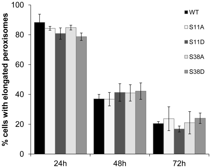
Pex11 proteins are involved in membrane elongation and division processes associated with the multiplication of peroxisomes. Human Pex11pβ has recently been linked to a new disorder affecting peroxisome morphology and dynamics. Here, we have analyzed the exact membrane topology of Pex11pβ. Studies with an epitope-specific antibody and protease protection assays show that Pex11pβ is an integral membrane protein with two transmembrane domains flanking an internal region exposed to the peroxisomal matrix and N- and C-termini facing the cytosol. A glycine-rich internal region within Pex11pβ is dispensable for peroxisome membrane elongation and division. However, we demonstrate that an amphipathic helix (Helix 2) within the first N-terminal 40 amino acids is crucial for membrane elongation and self-interaction of Pex11pβ. Interestingly, we find that Pex11pβ self-interaction strongly depends on the detergent used for solubilization. We also show that N-terminal cysteines are not essential for membrane elongation, and that putative N-terminal phosphorylation sites are dispensable for Pex11pβ function. We propose that self-interaction of Pex11pβ regulates its membrane deforming activity in conjunction with membrane lipids.


Conflict of interest statement

Competing Interests: The authors have declared that no competing interests exist.

Figures

Figure 1
Figure 1. Permeabilization of the peroxisome membrane is required for epitope recognition of the Pex11pβ antibody.
COS-7 cells were transfected with Pex11pβ-Myc (A–I) or YFP-Pex11pβ (J–R) and fixed after 24 hours. Cell membranes were permeabilized with 0.2% Triton X-100 (TX-100) (A–C, J–L), 25 µg/ml digitonin (D–F, M–O) or methanol (MetOH) (G–I, P–R) prior to immunostaining with anti-Myc (A, D, G) and anti-Pex11pβ (middle column) antibodies. Note that Pex11pβ-Myc is liberated from peroxisomal membranes after postfixation TX-100 treatment (A–C), while YFP-Pex11pβ is retained (J–L). Bars, 20 µm.
Figure 2
Figure 2. HsPex11pβ is an integral membrane protein with two transmembrane spans flanking a protease-protected region.
(A) COS-7 cells were transfected with Myc-tagged Pex11pα, Pex11pβ, or Pex11pγ and subjected to carbonate extraction (Carb.) at pH 11.5 or were mock treated (Con). Equal amounts of protein (P, membrane fraction; S, carbonate extract) were separated by SDS-PAGE on 12.5% acrylamide gels and subjected to immunoblotting with anti-Myc antibodies. PMP70 and Pex19p served as controls for integral and peripheral proteins, respectively. (B) Schematic view of potential results of a proteinase K (PK) digest depending on the number and location of putative transmembrane spans within Pex11pβ (see also Fig. S2). AB, epitope recognized by anti-Pex11pβ. (C) COS-7 cells were transfected with YFP-Pex11pβ or mock transfected (UT). 48 h after transfection, peroxisome-enriched fractions were prepared. Equal amounts of protein were digested with proteinase K in the presence or absence of Triton X-100 (TX-100). Controls were left untreated. Samples were separated by 12.5% SDS-PAGE and immunoblotted using anti-Pex11pβ. As a loading control, the membrane was re-incubated with anti-GFP after membrane stripping. Asterisks indicate the YFP-Pex11pβ band before and after digest. Note that the nonspecific band (approx. 60 kDa) is no longer recognized after repeated use of the Pex11pβ antibody (see D). (D) As an alternative to (C), peroxisome fractions were ruptured by sonication prior to proteinase K digest and immunoblotted as described. As a loading control, the membrane was re-incubated with anti-GFP. Successful membrane rupture was verified by incubation with anti-AOX, a peroxisomal matrix marker.
Figure 3
Figure 3. A glycine-rich internal region specific for human Pex11pβ is dispensable for peroxisome elongation and division.
COS-7 cells expressing Myc-Pex11pβ (A, C) and Myc-Pex11pβΔGly (B, D) were processed for immunofluorescence microscopy after 12 and 48 h using anti-Myc (A–D). (E) Quantitative evaluation of peroxisome morphology over time. Data are from 3 independent experiments and are presented as means ± S.D. Bars, 20 µm.
Figure 4
Figure 4. Phospho-mimicking mutants of Pex11pβ do not influence peroxisomal elongation and division.
COS-7 cells expressing Pex11pβ-Myc, Pex11pβ-MycS11A, Pex11pβ-MycS11D, Pex11pβ-MycS38A and Pex11pβ-MycS38D were processed for immunofluorescence using anti-Myc and anti-Pex14p antibodies (Suppl. Fig. S4) and peroxisome morphology was quantified. Data are from 3 independent experiments and are presented as means ± S.D.
Figure 5
Figure 5. An intact Helix 2 within the first 40 N-terminal aa of Pex11pβ is required to elongate the peroxisomal membrane.
COS-7 cells were transfected with Pex11pβ-Myc (A–C), the N-terminal deletions Pex11pβΔN40-Myc (D–F), Pex11pβΔN60-Myc, Pex11pβΔN70-Myc and the Helix 2-breaking mutant Pex11pβ-MycA21P. Cells were processed for immunofluorescence microscopy after 24 h using anti-Myc (A, D) and anti-PMP70 (B, E) antibodies. (G) Quantitative evaluation of peroxisome morphology. Data are from 3–4 independent experiments and are presented as means ± S.D. (*p<0.01). Bars, 20 µm.
Figure 6
Figure 6. ΔN40-Pex11pβ-Myc shows altered membrane distribution within tubular peroxisomal accumulations (TPAs) and fails to induce them when co-expressed with YFP-Pex11pβ.
COS-7 cells were co-transfected with Pex11pβ-YFP/Pex11pβΔN40-Myc (A–C), YFP-Pex11pβ/Pex11pβ-Myc (D–F), or YFP-Pex11pβ/Pex11pβΔN40-Myc (G–I) and processed as described below. Note that Pex11pβ-YFP localizes to tubular membranes (A, C), whereas Pex11pβΔN40-Myc distributes over both tubular and spherical membrane domains (B, C). Note that N-terminally tagged YFP-Pex11pβ only induces TPAs when co-expressed with Pex11pβ-Myc (D–F), but not with Pex11pβΔN40-Myc (G–I). (J) Quantitative evaluation of TPA formation in cells expressing Pex11pβ-YFP (a strong inducer of TPAs), YFP-Pex11pβ or co-expressing YFP-Pex11pβ/Pex11pβ-Myc or YFP-Pex11pβ/Pex11pβΔN40-Myc. Cells were fixed after 24 h, stained for immunofluorescence with anti-Myc antibodies and analyzed. Data are from 3–4 independent experiments and are presented as means ± S.D. (*p<0.01). Bars, 20 µm.
Figure 7
Figure 7. An intact Helix 2 within the first 40 N-terminal aa of Pex11pβ influences dimer formation.
COS-7 cells expressing Pex11pβ-Myc (WT) (A, B), Pex11pβΔN40-Myc (B), or Pex11pβ-MycA21P (B) were fixed with 4% para-formaldehyde 24 h after transfection and subjected to postfixation Triton X-100 (TX) or digitonin (Dig) extraction. Equal amounts of protein from supernatants (S) (TX-extracts), remaining cell pellets (P) and untreated lysates (L) were separated by 10% SDS-PAGE and immunoblotted using anti-Myc. Note that Pex11pβ-Myc is extracted by postfixation Triton X-100 treatment but not by digitonin (A). (C) Crosslinking of Pex11pβ-Myc with DSP. COS-7 cells expressing Pex11pβ-Myc were cross-linked with DSP and either lysed with 1% Triton X-100 or 1% digitonin. Equal protein amounts of the lysates were separated by reducing and non-reducing (non-red.) SDS-PAGE and immunoblotted using anti-Myc. Arrowheads highlight monomeric and dimeric forms of Pex11pβ-Myc. (D) Migration of Pex11pβ in native sucrose gradients. COS-7 cells expressing Pex11pβ-Myc were either lysed in buffer containing 1% Triton X-100 (after cross-linking with DSP) (TX, CL) or in buffer containing 1% digitonin (without cross-linking) (Dig). Cell lysates were applied on top of each gradient (*), separated by sucrose density gradient ultracentrifugation (10–47%) into 12 fractions and analyzed by immunoblotting using anti-Myc. A gradient with a molecular mass marker was run in parallel for size calibration; correspondent masses are indicated at the bottom. Note the difference in the molecular mass of Pex11pβ complexes indicating different oligomerization states depending on the detergent used.
Figure 8
Figure 8. Pex11pβ-induced peroxisomal division is impaired under lipid-free culture conditions.
COS-7 cells stably expressing a GFP-PTS1 fusion protein targeted to peroxisomes were incubated in lipid-free Panserin™ medium and transfected with Pex11pβ-Myc. Cells were processed for immunofluorescence using anti-Myc. Bar, 20 µm.
Figure 9
Figure 9. Mutations of N-terminal cysteines within Pex11pβ do not affect peroxisome membrane elongation.
COS-7 cells were transfected with Pex11pβ-Myc (A–C), Pex11pβ-MycC18S-C25S (D–F) and Pex11pβ-MycC18S-C25S-C85S (G–I), and were processed for immunofluorescence microscopy 24 h after transfection using anti-Myc (A, D, G) and anti-Pex14p (B, E, H) antibodies. (J) Quantitative evaluation of peroxisome morphology. Data are from 3 independent experiments and are presented as means ± S.D. Bars, 20 µm.

Similar articles

Cited by

References

    1. Wanders RJ, Waterham HR (2006) Peroxisomal disorders: the single peroxisomal enzyme deficiencies. Biochim Biophys Acta 1763: 1707–1720. - PubMed
    1. Dixit E, Boulant S, Zhang Y, Lee AS, Odendall C, et al. (2010) Peroxisomes are signaling platforms for antiviral innate immunity. Cell 141: 668–681. - PMC - PubMed
    1. Ivashchenko O, Van Veldhoven PP, Brees C, Ho YS, Terlecky SR, et al. (2011) Intraperoxisomal redox balance in mammalian cells: oxidative stress and interorganellar cross-talk. Mol Biol Cell 22: 1440–1451. - PMC - PubMed
    1. Islinger M, Grille S, Fahimi HD, Schrader M (2012) The peroxisome: an update on mysteries. Histochem Cell Biol 137: 547–574. - PubMed
    1. Lizard G, Rouaud O, Demarquoy J, Cherkaoui-Malki M, Iuliano L (2012) Potential Roles of Peroxisomes in Alzheimer's Disease and in Dementia of the Alzheimer's Type. J Alzheimers Dis 29: 241–254. - PubMed

Publication types

LinkOut - more resources