

Molecular and functional evidence of HCN4 and caveolin-3 interaction during cardiomyocyte differentiation from human embryonic stem cells
- PMID: 23311301
- PMCID: PMC3657289
- DOI: 10.1089/scd.2012.0247
Molecular and functional evidence of HCN4 and caveolin-3 interaction during cardiomyocyte differentiation from human embryonic stem cells
Abstract
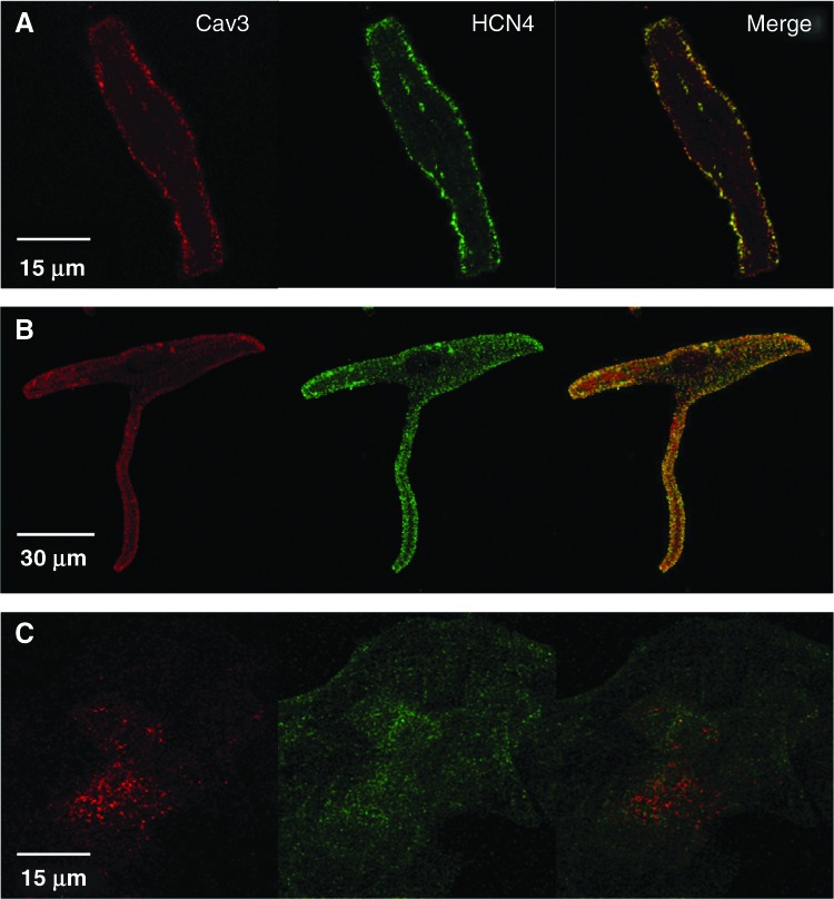
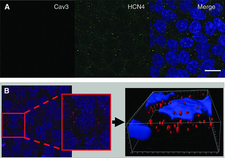
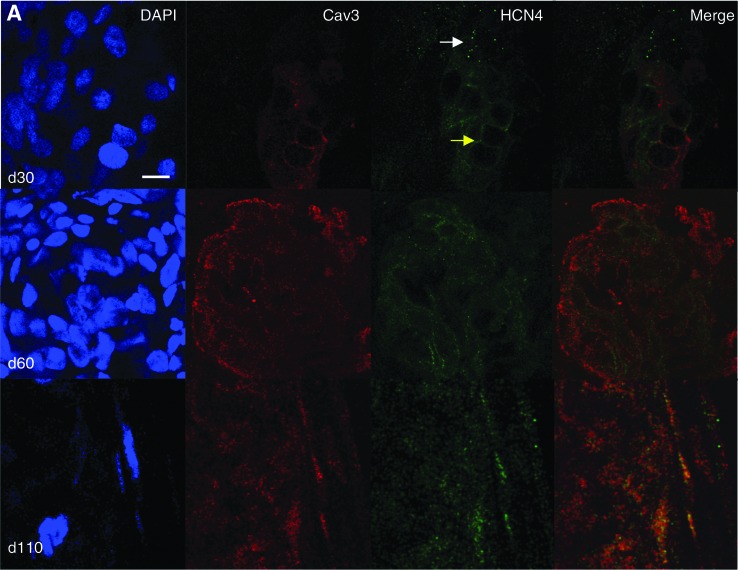
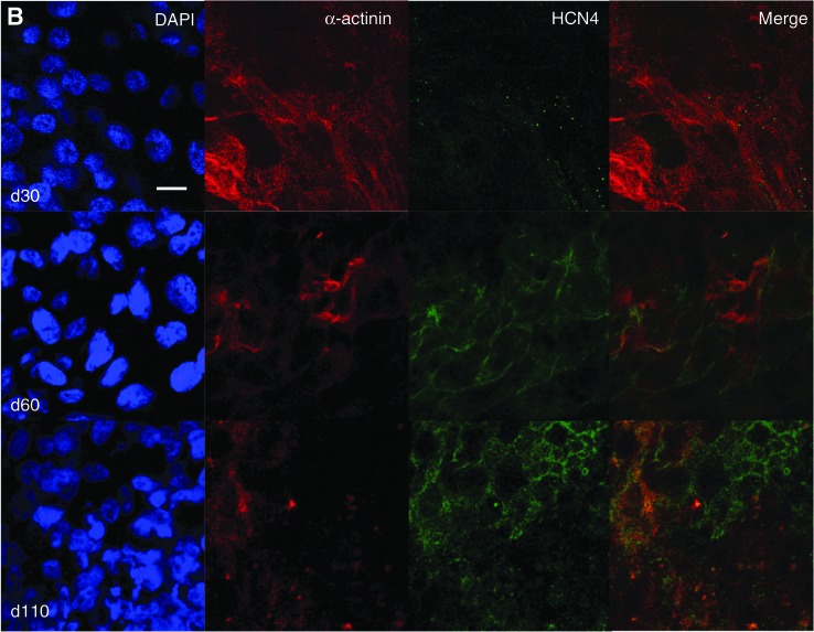
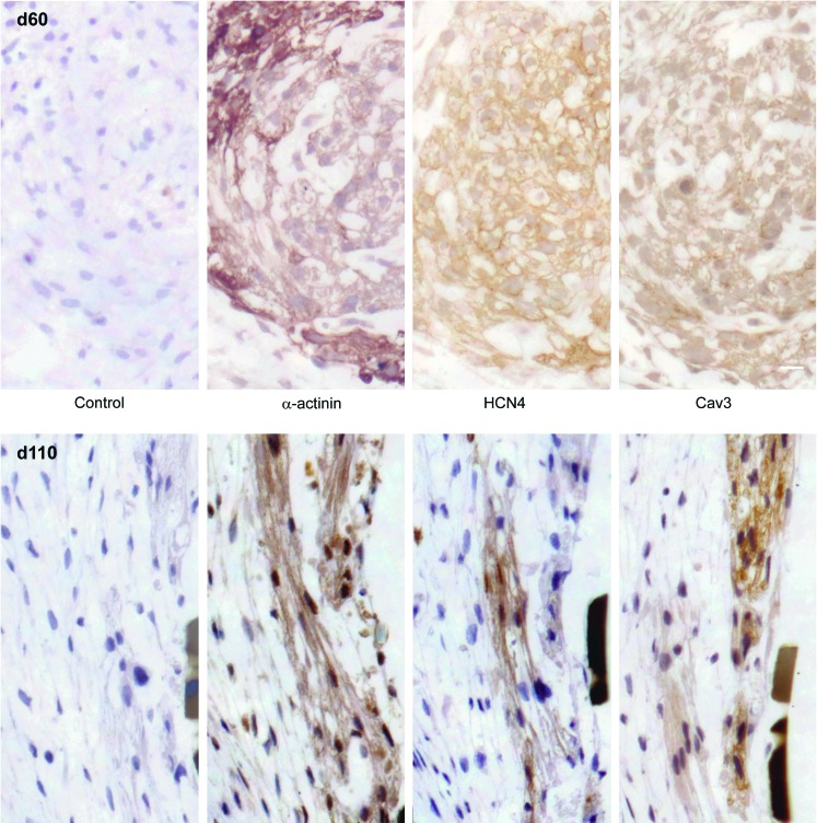
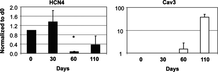
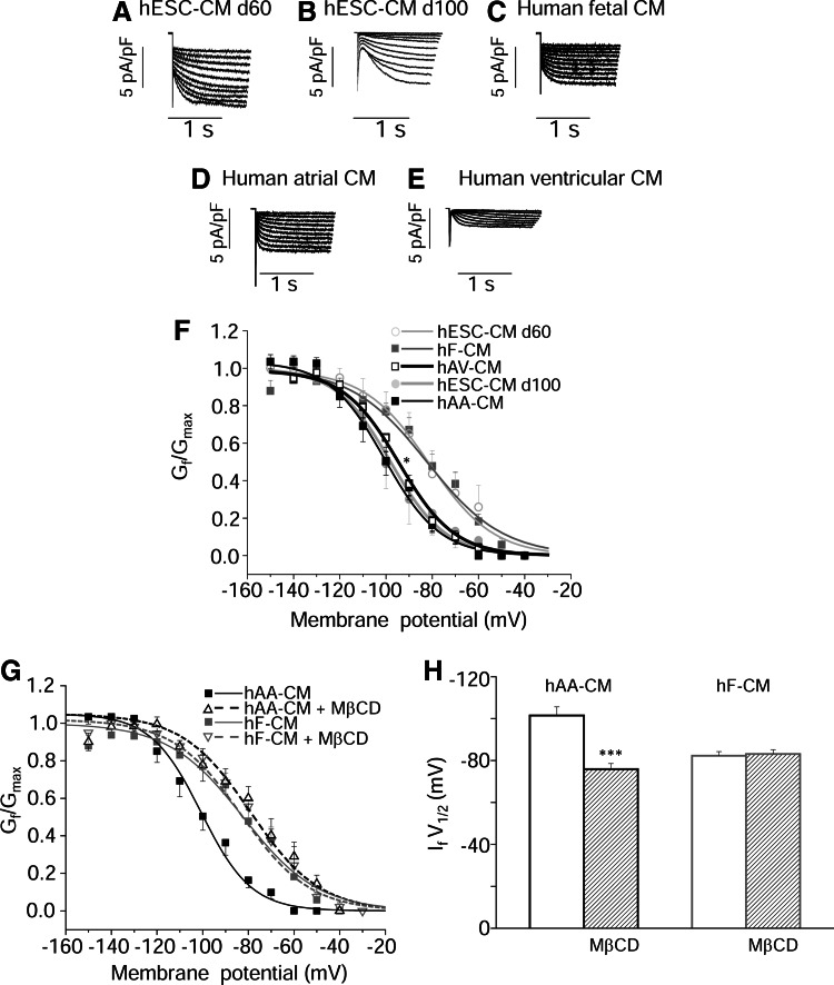
Maturation of human embryonic stem cell-derived cardiomyocytes (hESC-CM) is accompanied by changes in ion channel expression, with relevant electrophysiological consequences. In rodent CM, the properties of hyperpolarization-activated cyclic nucleotide-gated channel (HCN)4, a major f-channel isoform, depends on the association with caveolin-3 (Cav3). To date, no information exists on changes in Cav3 expression and its associative relationship with HCN4 upon hESC-CM maturation. We hypothesize that Cav3 expression and its compartmentalization with HCN4 channels during hESC-CM maturation accounts for the progression of f-current properties toward adult phenotypes. To address this, hESC were differentiated into spontaneously beating CM and examined at ∼30, ∼60, and ∼110 days of differentiation. Human adult and fetal CM served as references. HCN4 and Cav3 expression and localization were analyzed by real time PCR and immunocyto/histochemistry. F-current was measured in patch-clamped single cells. HCN4 and Cav3 colocalize in adult human atrial and ventricular CM, but not in fetal CM. Proteins and mRNA for Cav3 were not detected in undifferentiated hESC, but expression increased during hESC-CM maturation. At 110 days, HCN4 appeared to be colocalized with Cav3. Voltage-dependent activation of the f-current was significantly more positive in fetal CM and 60-day hESC-CM (midpoint activation, V1/2, ∼ -82 mV) than in 110-day hESC-CM or adult CM (V1/2∼-100 mV). In the latter cells, caveolae disruption reversed voltage dependence toward a more positive or an immature phenotype, with V1/2 at -75 mV, while in fetal CM voltage dependence was not affected. Our data show, for the first time, a developmental change in HCN4-Cav3 association in hESC-CM. Cav3 expression and its association with ionic channels likely represent a crucial step of cardiac maturation.
Figures
Similar articles
-
Caveolin-3 associates with and affects the function of hyperpolarization-activated cyclic nucleotide-gated channel 4.Biochemistry. 2008 Nov 25;47(47):12312-8. Biochemistry. 2008. PMID: 19238754 Free PMC article.
-
Non-cardiomyocytes influence the electrophysiological maturation of human embryonic stem cell-derived cardiomyocytes during differentiation.Stem Cells Dev. 2010 Jun;19(6):783-95. doi: 10.1089/scd.2009.0349. Stem Cells Dev. 2010. PMID: 20001453 Free PMC article.
-
Associated changes in HCN2 and HCN4 transcripts and I(f) pacemaker current in myocytes.Biochim Biophys Acta. 2009 May;1788(5):1138-47. doi: 10.1016/j.bbamem.2009.02.011. Epub 2009 Feb 21. Biochim Biophys Acta. 2009. PMID: 19236845 Free PMC article.
-
The structure of the apo cAMP-binding domain of HCN4 - a stepping stone toward understanding the cAMP-dependent modulation of the hyperpolarization-activated cyclic-nucleotide-gated ion channels.FEBS J. 2018 Jun;285(12):2182-2192. doi: 10.1111/febs.14408. Epub 2018 Mar 14. FEBS J. 2018. PMID: 29444387 Review.
-
Insights into cardiac conduction system formation provided by HCN4 expression.Trends Cardiovasc Med. 2015 Jan;25(1):1-9. doi: 10.1016/j.tcm.2014.08.009. Epub 2014 Sep 6. Trends Cardiovasc Med. 2015. PMID: 25442735 Free PMC article. Review.
Cited by
-
Human iPSC modelling of a familial form of atrial fibrillation reveals a gain of function of If and ICaL in patient-derived cardiomyocytes.Cardiovasc Res. 2020 May 1;116(6):1147-1160. doi: 10.1093/cvr/cvz217. Cardiovasc Res. 2020. PMID: 31504264 Free PMC article.
-
Damage-inducible intragenic demethylation of the human TP53 tumor suppressor gene is associated with transcription from an alternative intronic promoter.Mol Carcinog. 2016 Dec;55(12):1940-1951. doi: 10.1002/mc.22441. Epub 2015 Dec 16. Mol Carcinog. 2016. PMID: 26676339 Free PMC article.
-
A detailed characterization of the hyperpolarization-activated "funny" current (If) in human-induced pluripotent stem cell (iPSC)-derived cardiomyocytes with pacemaker activity.Pflugers Arch. 2021 Jul;473(7):1009-1021. doi: 10.1007/s00424-021-02571-w. Epub 2021 May 2. Pflugers Arch. 2021. PMID: 33934225 Free PMC article.
-
Absence of full-length dystrophin impairs normal maturation and contraction of cardiomyocytes derived from human-induced pluripotent stem cells.Cardiovasc Res. 2020 Feb 1;116(2):368-382. doi: 10.1093/cvr/cvz109. Cardiovasc Res. 2020. PMID: 31049579 Free PMC article.
-
Toward an in vitro human pacemaker.Pflugers Arch. 2021 Jul;473(7):989-990. doi: 10.1007/s00424-021-02585-4. Epub 2021 May 25. Pflugers Arch. 2021. PMID: 34032889 No abstract available.
References
-
- Barbuti A. Gravante B. Riolfo M. Milanesi R. Terragni B. DiFrancesco D. Localization of pacemaker channels in lipid rafts regulates channel kinetics. Circ Res. 2004;94:1325–1331. - PubMed
-
- Barbuti A. Scavone A. Mazzocchi N. Terragni B. Baruscotti M. Difrancesco D. A caveolin-binding domain in the HCN4 channels mediates functional interaction with caveolin proteins. J Mol Cell Cardiol. 2012;53:187–195. - PubMed
-
- Sartiani L. Bettiol E. Stillitano F. Mugelli A. Cerbai E. Jaconi ME. Developmental changes in cardiomyocytes differentiated from human embryonic stem cells: a molecular and electrophysiological approach. Stem Cells. 2007;25:1136–1144. - PubMed
-
- Cerbai E. Mugelli A. I(f) in non-pacemaker cells: role and pharmacological implications. Pharmacol Res. 2006;53:416–423. - PubMed
Publication types
MeSH terms
Substances
LinkOut - more resources
Full Text Sources
Other Literature Sources
