Camouflage treatment of skeletal Class III malocclusion with multiloop edgewise arch wire and modified Class III elastics by maxillary mini-implant anchorage
- PMID: 23311602
- PMCID: PMC8754039
- DOI: 10.2319/091312-730.1
Camouflage treatment of skeletal Class III malocclusion with multiloop edgewise arch wire and modified Class III elastics by maxillary mini-implant anchorage
Abstract
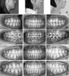
Objective: To evaluate the effect of the multiloop edgewise arch wire (MEAW) technique with maxillary mini-implants in the camouflage treatment of skeletal Class III malocclusion.
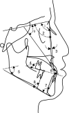
Materials and methods: Twenty patients were treated with the MEAW technique and modified Class III elastics from the maxillary mini-implants. Twenty-four patients were treated with MEAW and long Class III elastics from the upper second molars as control. Lateral cephalometric radiographs were obtained and analyzed before and after treatment, and 1 year after retention.
Results: Satisfactory occlusion was established in both groups. Through principal component analysis, it could be concluded the anterior-posterior dental position, skeletal sagittal and vertical position, and upper molar vertical position changed within groups and between groups; vertical lower teeth position and Wits distance changed in the experimental group and between groups. In the experimental group, the lower incisors tipped lingually 2.7 mm and extruded 2.4 mm. The lingual inclination of the lower incisors increased 3.5°. The mandibular first molars tipped distally 9.1° and intruded 0.4 mm. Their cusps moved 3.4 mm distally. In the control group, the upper incisors proclined 3°, and the upper first molar extruded 2 mm. SN-MP increased 1.6° and S-Go/N-ME decreased 1.
Conclusions: The MEAW technique combined with modified Class III elastics by maxillary mini-implants can effectively tip the mandibular molars distally without any extrusion and tip the lower incisors lingually with extrusion to camouflage skeletal Class III malocclusions. Clockwise rotation of the mandible and further proclination of upper incisors can be avoided. The MEAW technique and modified Class III elastics provided an appropriate treatment strategy especially for patients with high angle and open bite tendency.
Figures


References
-
- Massler M, Frankel JM. Prevalence of malocclusion in children aged 14 to 18 years. Am J Orthod. 1951;37:751–768. - PubMed
-
- Thilander B, Myrberg N. The prevalence of malocclusion in Swedish schoolchildren. Scand J Dent Res. 1973;81:12–21. - PubMed
-
- Irie M, Nakamura S. Orthopedic approach to severe skeletal Class III malocclusion. Am J Orthod. 1975;67:377–392. - PubMed
-
- Baccetti T, Reyes BC, McNamara JA., Jr Gender differences in Class III malocclusion. Angle Orthod. 2005;75:510–520. - PubMed
-
- Cozza P, Marino A, Mucedero M. An orthopaedic approach to the treatment of Class III malocclusions in the early mixed dentition. Eur J Orthod. 2004;26:191–199. - PubMed
Publication types
MeSH terms
Substances
LinkOut - more resources
Full Text Sources
Other Literature Sources

