Review
. 2013 Aug 20;19(6):595-637.
doi: 10.1089/ars.2012.4785. Epub 2013 Mar 6.

Redox control of inflammation in macrophages

Affiliations
Review

Redox control of inflammation in macrophages

Bernhard Brüne et al. Antioxid Redox Signal. .

Abstract

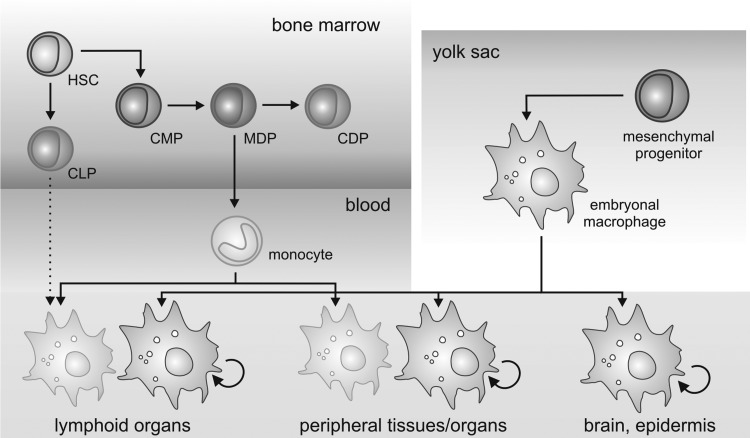
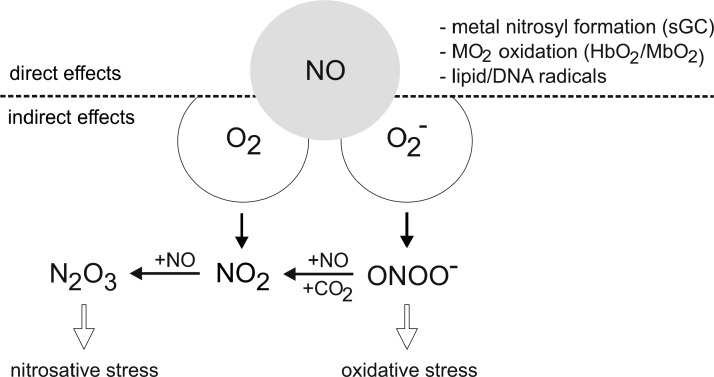
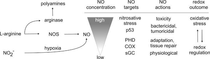
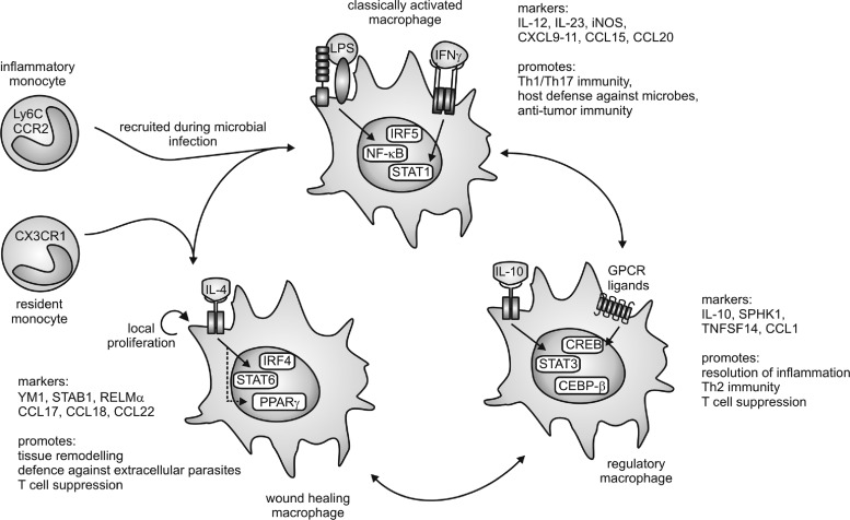
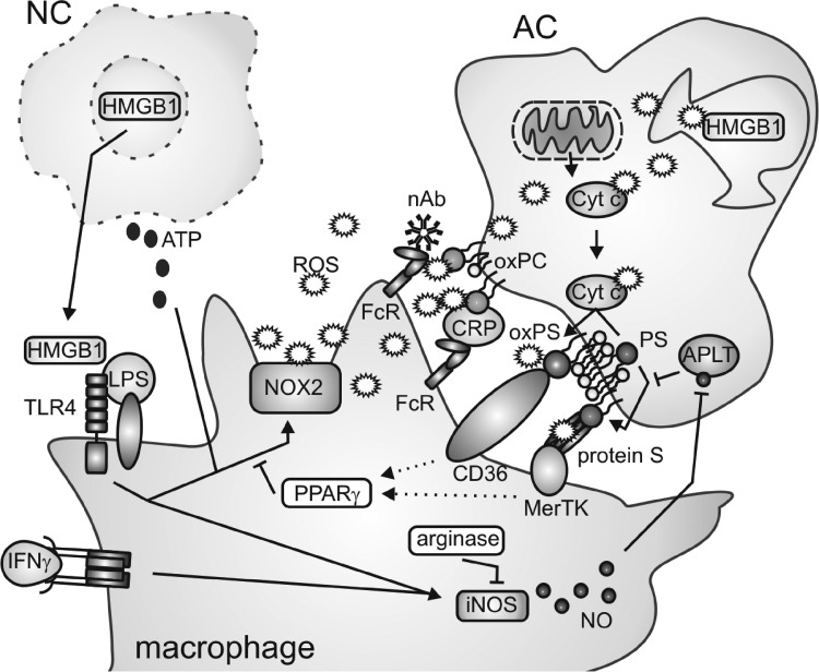
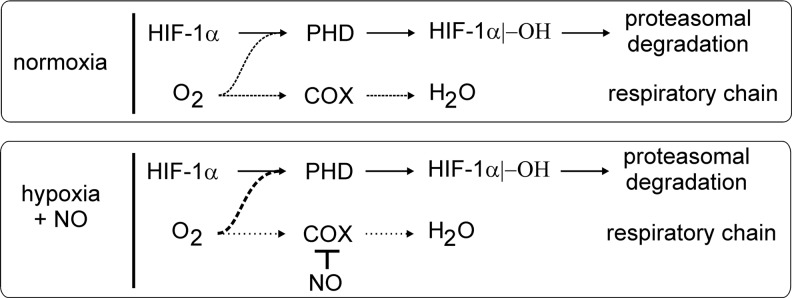
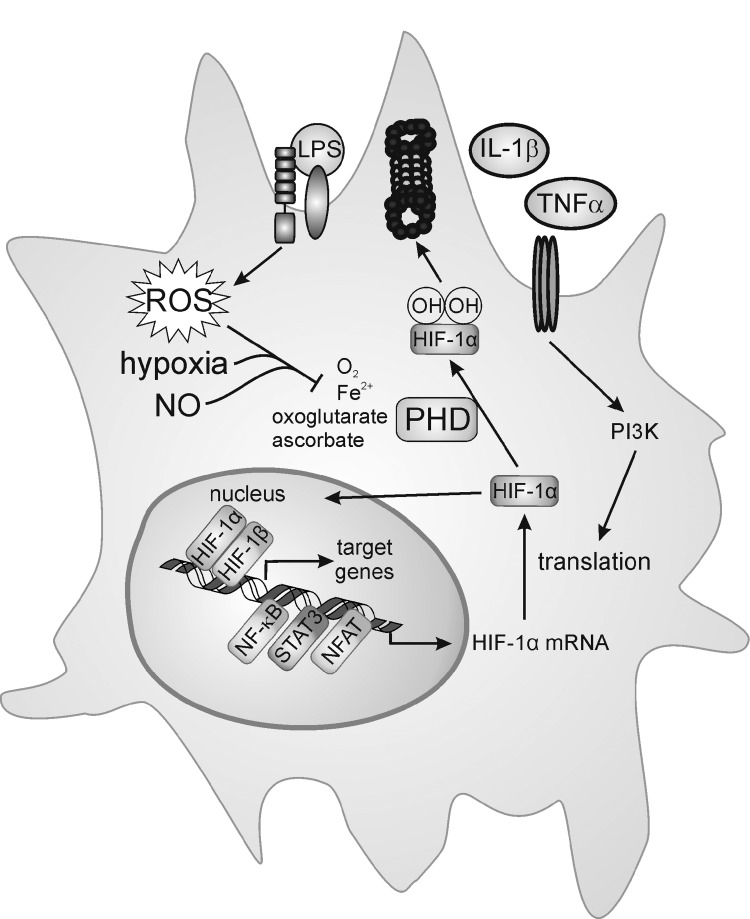
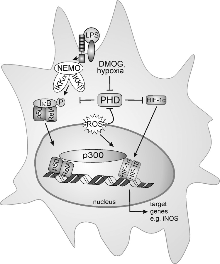
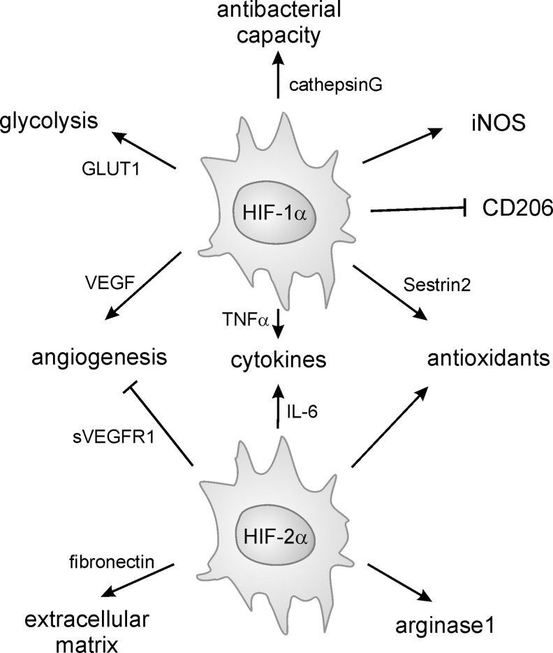
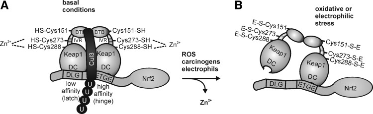
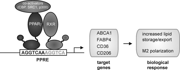
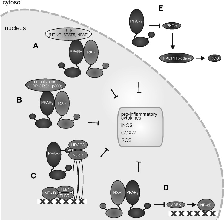
Macrophages are present throughout the human body, constitute important immune effector cells, and have variable roles in a great number of pathological, but also physiological, settings. It is apparent that macrophages need to adjust their activation profile toward a steadily changing environment that requires altering their phenotype, a process known as macrophage polarization. Formation of reactive oxygen species (ROS), derived from NADPH-oxidases, mitochondria, or NO-producing enzymes, are not necessarily toxic, but rather compose a network signaling system, known as redox regulation. Formation of redox signals in classically versus alternatively activated macrophages, their action and interaction at the level of key targets, and the resulting physiology still are insufficiently understood. We review the identity, source, and biological activities of ROS produced during macrophage activation, and discuss how they shape the key transcriptional responses evoked by hypoxia-inducible transcription factors, nuclear-erythroid 2-p45-related factor 2 (Nrf2), and peroxisome proliferator-activated receptor-γ. We summarize the mechanisms how redox signals add to the process of macrophage polarization and reprogramming, how this is controlled by the interaction of macrophages with their environment, and addresses the outcome of the polarization process in health and disease. Future studies need to tackle the option whether we can use the knowledge of redox biology in macrophages to shape their mediator profile in pathophysiology, to accelerate healing in injured tissue, to fight the invading pathogens, or to eliminate settings of altered self in tumors.


Figures

FIG. 1.
FIG. 1.
Developmental sources of adult tissue macrophages. Adult tissue macrophages are derived either from embryonic macrophages that develop early in the embryonal yolk sac and proliferate in situ, or from blood monocytes that develop along a specific line of progenitors in the bone marrow. There is speculation about a lymphoid source of macrophages in the lymphoid organs (dotted arrow). HSC, hematopoietic stem cell; CMP, common myeloid progenitor; CLP, common lymphoid progenitor; MDP, monocyte/dendritic cell progenitor; CDP, common dendritic cell progenitor. For details, see text.
FIG. 2.
FIG. 2.
Direct and indirect signaling of NO. Direct effects of NO are discriminated from indirect ones by the reactions with oxygen or superoxide radical anion, provoking oxidative or nitrosative stress. For details, see the text.
FIG. 3.
FIG. 3.
NO actions at distinct concentrations. Formation of NO by NO synthase (NOS) or derived from nitrate under hypoxia with NOS activity being antagonized by arginase, which consumes l-arginine as well. Exemplified are relative levels of NO required for distinct target interactions and accompanying biological outcomes ranging from redox regulation to oxidative stress. For details, see the text.
FIG. 4.
FIG. 4.
Macrophage subtypes. Macrophages can be polarized into three functional activation states. Stimulation with lipopolysaccharide (LPS) and interferon (IFN)γ generates classically activated macrophages that are characterized by expression of nuclear factor kappa light-chain enhancer of activated B-cells (NF-κB), signal transducer and activator of transcription 1 (STAT1), and interferon regulatory factor 5 (IRF5). Downstream of the interleukin-4 (IL-4) receptor, activation of STAT6, peroxisome proliferator-activated receptor (PPAR)γ, and IRF4 generate wound-healing macrophages. Regulatory macrophages are characterized by expression of STAT3, CCAAT/enhancer-binding protein (CEBP)-β, and cAMP-responsive element-binding protein (CREB) downstream of stimulation with IL-10, G-protein-coupled receptor (GPCR) ligands, and others. Arrows between the macrophage subtypes indicate convertibility as well as the occurrence of mixed or hybrid phenotypes. Markers of the distinct macrophage populations as well as their functional profiles are indicated. For details, see text. On top of polarization of mature macrophages, inflammatory versus resident monocytes may exhibit the potential to differentiate to classically activated or wound-healing macrophages, respectively. During Th1 inflammation, classically activated macrophages are initially recruited to the site of infection, whereas during Th2 inflammation, enhanced macrophage numbers may result from local proliferation.
FIG. 5.
FIG. 5.
Redox regulation of macrophage interaction with dying cells. Release of alarmins such as high-mobility group box 1 (HMGB1) and adenosine triphosphate (ATP) from necrotic cells (NC) induces macrophage activation concomitant with upregulation of inducible NO synthase (iNOS) and activation of the NOX2-containing NADPH oxidase complex. In apoptotic cells (AC), reactive oxygen species (ROS) generated at the mitochondria deactivate HMGB1 by oxidation. Cytochrome c (Cyt c) released from mitochondria during apoptosis in an oxidized form in turn oxidizes phosphatidylserine (PS) at the inner leaflet of the plasma membrane, leading to its externalization, which requires inhibition of aminophospholipid translocase. PS, oxPS, as well as oxidized phosphatidylcholine (oxPC) are recognized by macrophage scavenger receptors through bridging molecules, which may themselves undergo oxidative modifications. Downstream of AC recognition, unknown mechanisms (dotted arrows) activate PPARγ, which inhibits the NOX2 complex in macrophages. Mechanisms of arginase induction and induction or repression of iNOS upon AC–macrophage interactions are controversially discussed and therefore are not clearly indicated. For details, please see the text. FcR: Fc (antibody) receptor; nAb: natural (IgM) antibody; CRP: C-reactive protein.
FIG. 6.
FIG. 6.
Redox biology of prolyl hydroxylases (PHD). ROS generated by NOX, an altered antioxidant status or mitochondrial respiration, as well as hypoxia or NO block PHD activity. This stabilizes hypoxia-inducible factor (HIF)-1α, allows the interaction with HIF-1β, and subsequent target gene activation. iNOS as one HIF-1-regulated target may start a positive amplification loop to stabilize HIF-1α. iNOS not only is an HIF-1 target but also a classical NF-κB-responsive gene, activated in LPS-stimulated macrophages. o- - -o; refers to the PHD and HIF-α interaction.
FIG. 7.
FIG. 7.
PHD activity and mitochondrial oxygen consumption. In the presence of oxygen, the PHD activity continuously hydroxylates HIF-1α, which allows its subsequent proteasomal degradation. Oxygen is also consumed during mitochondrial respiration, with cytochrome-c oxidase (COX) reducing oxygen to water. Inhibition of COX by NO shunts the mitochondrial-used oxygen toward PHD activity. For details, see the text.
FIG. 8.
FIG. 8.
Transcriptional, translational, and post-translational regulation of HIF-1α. In the presence of oxygen, PHDs continuously hydroxylate HIF-1α protein and thereby mark it for proteasomal degradation. Hypoxia, NO, or ROS inhibit PHD activity to stabilize HIF-1α, which dimerizes with HIF-1β to promote target gene induction. IL-1β, tumor necrosis factor-α (TNFα), or LPS increases HIF-1α translation or use ROS to block PHD activity, while inflammatory-associated transcription factors such as NF-κB, STAT3, and/or nuclear factor of activated T-cells (NFAT) increase HIF-1α mRNA expression. For details, see the text.
FIG. 9.
FIG. 9.
Regulation of NF-κB and HIF-1α by hypoxia. Proinflammatory macrophage activation by LPS causes formation of the I-kappa-B kinase (IKK) complex composed of inhibitor of NF-κB (IκB) kinase g (NEMO), IκB kinase α (IKKa), and IκB kinase β (IKKb), which then phosphorylates IκB to release the NF-κB dimer p50 and RelA. PHDs hydroxylate and thereby inactivate IKKβ. Under hypoxia, or by blocking PHD activity with dimethyl oxalylglycine (DMOG), NF-κB is activated, and HIF-1α is stabilized. Both transcription factors translocate to the nucleus, bind to the DNA, and recruit cofactors such as E1A-binding protein p300 (p300/CBP) to induce, at least in part, the overlapping target genes, that is, iNOS. ROS in activated macrophages attenuates PHD activity, but additionally increase binding of p300/CBP to HIF-1. For details, see the text.
FIG. 10.
FIG. 10.
Impact of HIF-1α and HIF-2α on macrophage polarization. HIF-1 and HIF-2 share, in part, similar targets, but the individual knockout of the HIF-α subunits in macrophages provokes different phenotypes. HIF-1 enhances glycolysis, the antibacterial capacity, including iNOS, but decreases expression of CD206. Although antioxidant enzymes are HIF-2 regulated, sestrin2 is an HIF-1 target in macrophages. Cytokine production is attenuated by the knockdown of both isoforms. While TNFα predominantly is under the influence of HIF-1, IL-6 is HIF-2 dominated. Angiogenesis is affected by HIF-1 (via vascular endothelial growth factor [VEGF]), while HIF-2 antagonizes angiogenesis (VEGF receptor1 [VEGFR1]). For details, see the text.
FIG. 11.
FIG. 11.
Mechanisms of nuclear erythroid-2-p45-related factor 2 (Nrf2) accumulation. Under basal conditions, (A) Nrf2 binds to its cytosolic inhibitor Kelch-like ECH-associated protein 1 (Keap1)/INrf2, an adaptor protein to the cullin-3 (Cul3) E3 ubiquitin ligase complex, which targets Nrf2 for proteasomal degradation. After oxidative, nitrosative, or electrophilic stress, (B) Keap1 releases Cul3 and Nrf2, which stabilizes Nrf2, with the subsequent binding of DnaJ homolog 1 (DJ1). This complex mediates the nuclear import of Nrf2. Protein kinase C (PKC) conferred Nrf2 phosphorylation at serine 40 (S40) to dissociate Keap1 and Cul3 from Nrf2, followed by its nuclear import with DJ-1 (C). In the nucleus, Nrf2 binds to antioxidant element-binding sites (D) and induces the expression of defensive genes after coassociation to small musculoaponeurotic fibrosarcoma oncogene homologues, activating transcription factor (ATF)-4, or JunD, as well as to the transcriptional coactivator p300/CBP. Nuclear Nrf2 is exported from the nucleus after GSK-3β-dependent Fyn phosphorylation (E), which itself translocates to the nucleus to phosphorylate Nrf2 at tyrosine568 (Y568). In the cytosol, Nrf2 is again degraded via Keap1 (A).
FIG. 12.
FIG. 12.
Hinge-and-latch model of Keap1 binding to Nrf2. (A) Under basal conditions, Keap1 forms a homodimer via their broad complex, tram–track and bric-a-brac domains (BTB). This domain contains one out of three cysteine residues (Cys151), which are important for the redox modification of Keap1. In the intervening region (IVR), two other relevant cysteine residues are located (Cys273 and Cys288). The sulfhydryl groups (–SH) of these cysteine residues remain unaltered and partially coordinate Zn2+. One Keap1 molecule of the homodimer binds via its double glycine repeat and the C-terminal region domain (DC) to the conserved amino acid motif aspartic acid-leucine-glycine (DLG) of Nrf2 with low affinity (latch). The other Keap molecule binds with its DC domain to the conserved amino acid motif glutamic acid-threonine-glycine-glutamic acid (ETGE) of Nrf2 with high affinity (hinge). (B) After oxidative, carcinogenic, or electrophilic stress, the three –SH groups (Cys151, Cys 273, and Cys288) are oxidatively modified (-S-E). This releases Keap1 from the Nrf2 DLG motif, whereas binding to the Nrf2 ETGE motif remains intact. However, Cul3 association is attenuated, thus stabilizing Nrf2.
FIG. 13.
FIG. 13.
PPARγ-dependent transactivation of target genes. PPARγ binds as a heterodimer with retinoid x receptor (RXR) to PPAR-responsive elements (PPRE) in the promoter regions of target genes. After coactivator binding, expression of target genes is induced. Biological outcomes are indicated. For details, see the text.
FIG. 14.
FIG. 14.
Anti-inflammatory roles of PPARγ not requiring DNA binding. PPARγ binds a heterodimer with RXR to proinflammatory transcription factors (A) and transcriptional coactivators (B). This binding to PPARγ/RXR attenuates an anti-inflammatory macrophage phenotype. Moreover, PPARγ prevents NF-κB expression by inhibiting nuclear receptor corepressor (NCoR) degradation. This repressor remains bound to DNA by SUMOylated PPARγ (C), blocking proinflammatory gene expression. By a so-far unknown mechanism, the PPARγ/RXR heterodimer attenuates mitogen-activated protein kinase (MAPK) activation (D), to block the downstream transcription factors. Cytosolic PPARγ binds to PKCα to inhibit its activation and translocation to the cell membrane, subsequently blocking NOX activation and ROS formation (E).
FIG. 15.
FIG. 15.
Role of Nrf2 and PPARγ in macrophage polarization. Classical activation of macrophages activates NOX2 via PKCα. ROS may kill bacteria or modify Keap1 to release Nrf2 and to activate the protective genes. Under conditions of high ROS production, PPARγ likely is redox modified (RM). This blocks transcription of PPARγ target genes, but does not interfere with its DNA-independent actions (removal of transcription factors and/or coactivators). In alternatively activated macrophages, ROS formation is attenuated by PPARγ-dependent PKCα inhibition, and Nrf2 is degraded. Redox modifications of PPARγ are reversed, allowing to regain transcription of genes such as CD36 or CD206.
FIG. 16.
FIG. 16.
ROS in endoplasmic reticulum (ER) stress-induced cell death. ER stress induces an unfolded protein response (UPR), enhancing transcriptional activation of C/EBP-homologous protein (CHOP) and its target ER oxidation 1α (ERO1α). ERO1α activates IP3 receptor 1 (IP3R1) and calcium release, which activates the calcium-/calmodulin-dependent protein kinase IIγ (CAMKIIγ)-JNK cascade and transcriptional upregulation of NOX2. NOX2 is also activated in an acidic compartment by signals derived from activation of pattern recognition receptors such as Toll-like receptor (TLR)2, heterodimerizing with scavenger receptor CD36, and activation of extracellular signal regulated kinase(s). NOX2-derived ROS further activate CAMKIIγ-JNK, to enhance the UPR signal, thus contributing to the mitochondrial membrane permeabilization and induction of apoptosis. The CAMKIIγ-JNK cascade also induces Fas transcription, activating the extrinsic apoptotic pathway.
FIG. 17.
FIG. 17.
ROS and NOD-like receptor family, pyrin domain containing 3 (NLRP3) inflammasome activation. TLR receptor agonists deliver a priming signal to increase IL-1β and NLRP3 transcription and pro-IL-1β production. Activating stimuli, for example, ATP, stimulate P2X7 receptor-mediated potassium efflux, which promotes mitochondrial ROS generation and oxidation of mitochondrial DNA (mtDNA). Oxidized mtDNA binds NLRP3 and drives the inflammasome assembly, consisting of oligomers of NLRP3, apoptosis-associated speck-like protein containing a CARD (ASC) and pro-caspase-1, culminating in caspase-1 cleavage, and activation and IL-1β processing. Lysosomal rupture and cathepsin release induced by cholesterol or monosodium urate crystals also activate the inflammasome. Damaged, ROS-producing mitochondria are removed by mitophagy, while inhibition of mitophagy by saturated fatty acids (SFA) increases mtROS and enhances inflammasome activation. ROS also support a priming signal.
FIG. 18.
FIG. 18.
Role of iron in macrophage polarization. Role of iron in controlling macrophage immune responses associated with changes in iron transport, storage, recycling, and acquisition. For details, see the text.
FIG. 19.
FIG. 19.
Role of lipocalin-2 (Lcn-2) as an alternative iron transporter. Signals generated by tumor cells initiate Lcn-2 expression in macrophages, its loading with iron, and release. Together with iron release via ferroportin, macrophages provide iron to tumor cells, which is used by them to stimulate their survival and growth. For details, see the text.

References

    1. Adamson S. Leitinger N. Phenotypic modulation of macrophages in response to plaque lipids. Curr Opin Lipidol. 2011;22:335–342. - PMC - PubMed
    1. Alexander A. Cai SL. Kim J. Nanez A. Sahin M. MacLean KH. Inoki K. Guan KL. Shen J. Person MD. Kusewitt D. Mills GB. Kastan MB. Walker CL. ATM signals to TSC2 in the cytoplasm to regulate mTORC1 in response to ROS. Proc Natl Acad Sci U S A. 2010;107:4153–4158. - PMC - PubMed
    1. Andrews NC. Anemia of inflammation: the cytokine-hepcidin link. J Clin Invest. 2004;113:1251–1253. - PMC - PubMed
    1. Apetoh L. Ghiringhelli F. Tesniere A. Criollo A. Ortiz C. Lidereau R. Mariette C. Chaput N. Mira JP. Delaloge S. Andre F. Tursz T. Kroemer G. Zitvogel L. The interaction between HMGB1 and TLR4 dictates the outcome of anticancer chemotherapy and radiotherapy. Immunol Rev. 2007;220:47–59. - PubMed
    1. Ariel A. Serhan CN. New lives given by cell death: macrophage differentiation following their encounter with apoptotic leukocytes during the resolution of inflammation. Front Immunol. 2012;3:4. - PMC - PubMed

Publication types

Substances