. 2013 Jan 12:14:3.
doi: 10.1186/1471-2121-14-3.

mTOR direct interactions with Rheb-GTPase and raptor: sub-cellular localization using fluorescence lifetime imaging

Affiliations

mTOR direct interactions with Rheb-GTPase and raptor: sub-cellular localization using fluorescence lifetime imaging

Rahul B Yadav et al. BMC Cell Biol. .

Abstract

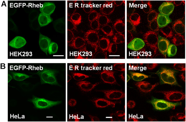
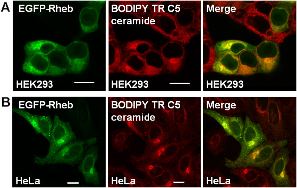
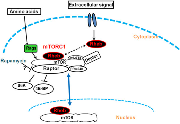
Background: The mammalian target of rapamycin (mTOR) signalling pathway has a key role in cellular regulation and several diseases. While it is thought that Rheb GTPase regulates mTOR, acting immediately upstream, while raptor is immediately downstream of mTOR, direct interactions have yet to be verified in living cells, furthermore the localisation of Rheb has been reported to have only a cytoplasmic cellular localization.

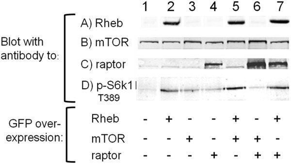
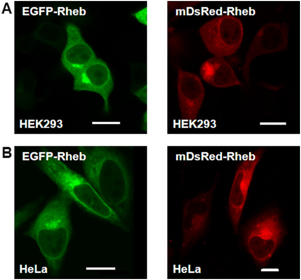
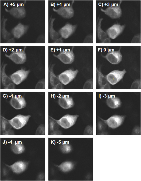
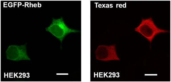
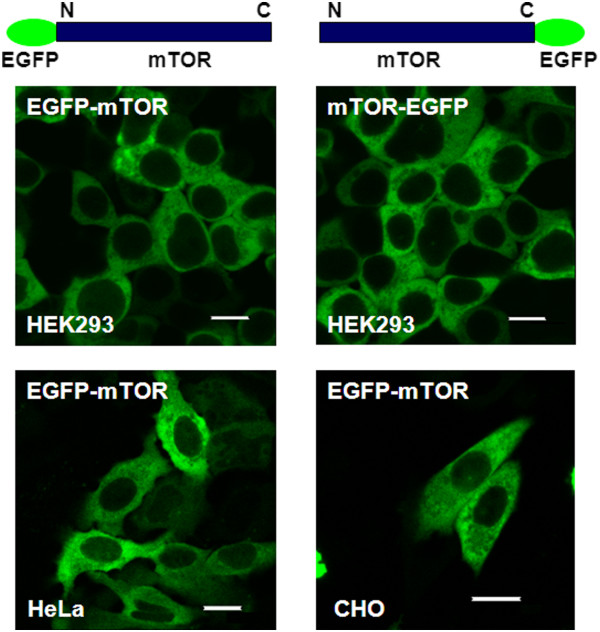
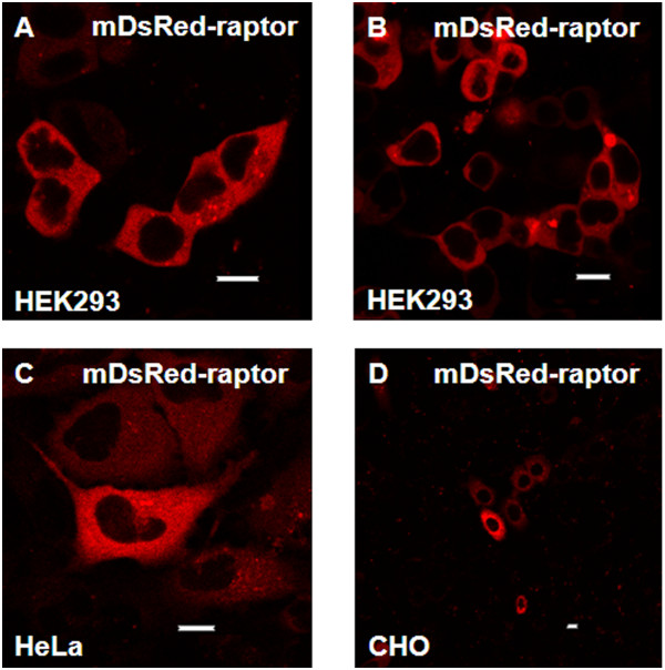
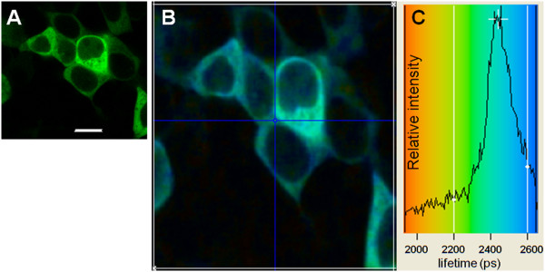
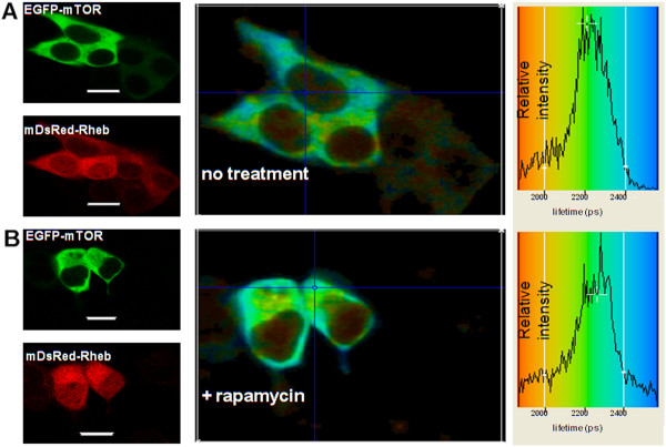
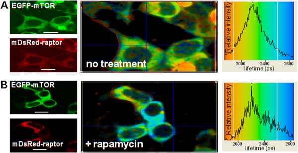
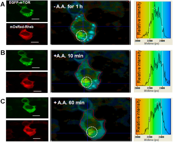
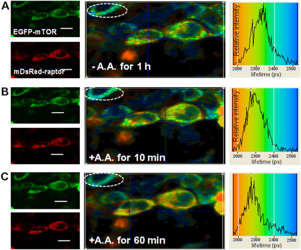
Results: In this study a cytoplasmic as well as a significant sub-cellular nuclear mTOR localization was shown , utilizing green and red fluorescent protein (GFP and DsRed) fusion and highly sensitive single photon counting fluorescence lifetime imaging microscopy (FLIM) of live cells. The interaction of the mTORC1 components Rheb, mTOR and raptor, tagged with EGFP/DsRed was determined using fluorescence energy transfer-FLIM. The excited-state lifetime of EGFP-mTOR of ~2400 ps was reduced by energy transfer to ~2200 ps in the cytoplasm and to 2000 ps in the nucleus when co-expressed with DsRed-Rheb, similar results being obtained for co-expressed EGFP-mTOR and DsRed-raptor. The localization and distribution of mTOR was modified by amino acid withdrawal and re-addition but not by rapamycin.

Conclusions: The results illustrate the power of GFP-technology combined with FRET-FLIM imaging in the study of the interaction of signalling components in living cells, here providing evidence for a direct physical interaction between mTOR and Rheb and between mTOR and raptor in living cells for the first time.


Figures

Figure 1
Figure 1
Functionality of EGFP-Rheb, EGFP-mTOR and DsRed-raptor determined by S6-kinase activity. EGFP-Rheb, EGFP-mTOR and DsRed-raptor were expressed in HEK cells and gels were run and blotted with the respective antibodies to Rheb, mTOR, raptor and S6-kinase (thr389). The expressed level of EGFP-Rheb is shown in Row A, lanes 2, 5 and 7 but the antibody was unable to pick up endogenous levels. The endogenous levels of mTOR can be seen in Row B, lanes 1, 2, 4 and 7 (with increased density when EGFP-mTOR is expressed (lanes 3, 5 and 6, high MW too close for large migration difference). The endogenous levels of raptor can be seen in Row C, lanes 1, 2, 3, and 5, with lanes 4, 6 and 7 showing DsRed-raptor expression.
Figure 2
Figure 2
EGFP-Rheb and DsRed-Rheb expression in mammalian cells. A) HEK293 cells and B) HeLa cells were transiently transfected with EGFP-Rheb (left panel) and DsRed-Rheb vector (right panel). 24h following transfection, and the live cells analyzed under a Nikon TE2000 U confocal microscope. Bar 8 μm.
Figure 3
Figure 3
3D Stack images of multiphoton-induced fluorescence of HEK293 expressing EGFP-Rheb. Fluorescence from TCSPC images of live HEK293 cells was acquired using multiphoton excitation (920 nm laser excitation, 520 nm emission) following 24h transfection. Raw data presented without further image processing. Images show clear Rheb nuclear localization. Yellow and red boxes (F) refer to nuclear and ER/Golgi regions taken for comparative photon count. Image size 120x100 μm.
Figure 4
Figure 4
Immunohistochemical staining of Rheb. HEK293 cells expressing EGFP-Rheb (LEFT PANEL) or cells fixed in 4% formaldehyde and treated with anti-Rheb antibody in conjugation with Texas red-labelled secondary antibody (RIGHT PANEL) were imaged with confocal microscopy. Bar 10 μm.
Figure 5
Figure 5
Sub-cellular localization of Rheb on ER. A) HEK293 cells; B) HeLa cells were transiently transfected with EGFP-Rheb. Twenty-four hours following transfection cells were stained with 1 μM ER Tracker red and live cells analyzed by confocal microscopy. The images reveal that Rheb localizes to the ER Bar 8 μm.
Figure 6
Figure 6
Sub-cellular localization of Rheb on Golgi. A) HEK293 cells; B) HeLa cells were transiently transfected with EGFP-Rheb. Twenty-four hours after transfection cells were stained with 5 μM BODIPY TR C5 ceramide and live cells analyzed by confocal microscopy. The images reveal that Rheb localizes to the Golgi. Bar 8 μm.
Figure 7
Figure 7
EGFP-mTOR expresses predominantly in the cytoplasm of mammalian cells. EGFP tagged mTOR expression was studied in HEK293, HeLa and CHO cells. The cells were transiently transfected with EGFP tagged mTOR and expression was analyzed in live cells following 48h of transfection using a Nikon TE2000 U inverted microscope and EC1 confocal system. (EGFP-mTOR; EGFP tagged at the N-terminal of mTOR and mTOR-EGFP; EGFP tagged at the C-terminal of mTOR). Bar 8 μm.
Figure 8
Figure 8
Sub-cellular localization of transiently transfected DsRed-raptor in different mammalian cell types. DsRed tagged raptor was expressed in HEK293 cells for A) 24h and B) 48h, HeLa cells for 48h, C) and CHO cells for 48h, D). Live cells were analyzed for various times following transfection using a Nikon TE2000 U confocal microscope. Bar 8 μm.
Figure 9
Figure 9
Fluorescence lifetime imaging of EGFP-mTOR expressed in HEK293 cells. A) Confocal image of EGFP-mTOR expressed in HEK293 cells following 48 h of transfection; B) Lifetime image of the same cells (colour coding for the lifetimes is shown in (C)), image size 120 x 110 μm; C) Lifetime distribution taken for the entire area (white border) shown in (B). The data collection time for lifetime images were optimally 3 accumulations of 30 sec. with an image dimension of 128×128 pixels (the blue cross lines are for single pixel lifetime values which were not used). A fluorescence lifetime value was determined from the distribution centre in (C), representing a value for the entire area shown in (B) and was ~2450 ± 100 ps, taken from three independent experiments.
Figure 10
Figure 10
DsRed-Rheb direct interaction with EGFP-mTOR in HEK293 cells and lack of effect of rapamycin. (A, B) LEFT PANELS: Confocal 488 nm excitation images of EGFP-mTOR (co-expressed with DsRed-Rheb, after 48 h of transfection) and 543 nm excitation images of DsRed-Rheb, without (A) and with (B) rapamycin (100 nM) treatment for 24 h. (A, B) MIDDLE PANELS: Lifetime images of the co-transfected cells (colour coding for the lifetimes is shown in the RIGHT PANELS). (A, B) RIGHT PANELS The lifetime distributions taken for the entire area (white border) as shown in the MIDDLE PANELS. The lifetime value from the data in (A) RIGHT PANEL for the EGFP (attached to mTOR) is reduced due by quenching by DsRed (attached to Rheb) from ~2450 ± 100 ps for EGFP alone (see Figure 9) to 2200 ± 100 ps (n=3) here, thus showing a direct interaction. The data in (B) RIGHT PANEL for the lifetime of the EGFP (attached to mTOR), after treatment with rapamycin there was no relief of the quenching indicating it does not impact on the direct interaction. The data collection time for lifetime images was optimally 3 accumulations of 30 sec. with an image dimension of 128×128 pixels, bar 8 μm.
Figure 11
Figure 11
EGFP-mTOR direct interaction with DsRed-raptor in HEK293 cells and lack of effect of rapamycin. (A, B) LEFT PANELS: Confocal 488 nm excitation images of EGFP-mTOR (co-expressed with DsRed-raptor, after 48 h of transfection) and 543 nm excitation images of DsRed-raptor, without (A) and with (B) rapamycin (100 nM) treatment for 24 h. (A, B) MIDDLE PANELS: Lifetime images of the co-transfected cells (colour coding for the lifetimes is shown in the RIGHT PANELS). (A, B) RIGHT PANELS The lifetime distributions taken for the the region taken here is within the area of the thin red line (not the entire area as previous Figures) as shown in the MIDDLE PANELS. The lifetime value from the data in (A) RIGHT PANEL for the EGFP (attached to mTOR) is reduced due to quenching by DsRed (attached to raptor) from ~2450 ± 100 ps for EGFP alone (see Figure 9) to 2300 ± 100 ps (n=3) here, thus showing a direct interaction. The data in (B) RIGHT PANEL for the lifetime of the EGFP (attached to mTOR) for the cell demarked by the thin red line (the cell below being omitted as it is not expressing DsRed-raptor, see LEFT PANEL), after treatment with rapamycin there was no effect on the lifetime centre (2300 ps ± 100 ps). The data collection time for lifetime images was optimally 3 accumulations of 30 sec. with an image dimension of 128×128 pixels, bar 8 μm.
Figure 12
Figure 12
Amino acid starvation and re-stimulation does not affect the direct interaction between EGFP-mTOR and DsRed-Rheb in perinuclear regions. (A-C) LEFT PANELS: Confocal 488 nm excitation images of EGFP-mTOR (co-expressed with DsRed-Rheb, after 48 h of transfection) and 543 nm excitation images of DsRed-Rheb, for HEK293 cells serum-starved overnight and then amino acid starved in D-PBS for 1 h (A) following which the amino acids were added back (B, C). Punctate structures appear after amino acid removal and disappear after adding back the amino acid and appear not to contain Rheb. A-C) MIDDLE PANELS, Lifetime images of the co-transfected cells (colour coding for the lifetimes is shown in the RIGHT PANELS). (A-C) RIGHT PANELS The lifetime distributions taken for the cell area (within red line) as shown in the MIDDLE PANELS. A, revealing both quenched and unquenched EGFP reflecting the mixture of perinuclear mTOR/Rheb and punctate mTOR), as reflected in the lifetime image; B) the same cells after 10 min of amino acid re-stimulation; C) after 60 minutes of amino acid re-stimulation when the punctate structures had disappeared. At all time-points the area marked by the white circle, mainly ER/Golgi, shows a quenched lifetime i.e. Rheb interacting with mTOR. Bar 8 μm.
Figure 13
Figure 13
Amino acid starvation and re-stimulation effect on the direct interaction between EGFP-mTOR and DsRed-raptor. (A-C) LEFT PANELS: Confocal 488 nm excitation images of EGFP-mTOR (co-expressed with DsRed-raptor, after 48 h of transfection) and 543 nm excitation images of DsRed-raptor, for HEK293 cells serum-starved overnight and then amino acid starved in D-PBS for 1 h (A) following which the amino acids were added back (B, C). Punctate structures appear after amino acid removal and disappear after adding back the amino acid. (A-C) MIDDLE PANELS, Lifetime images of the co-transfected cells. The colour coding for the lifetime is shown in the right panel which is a lifetime distribution of the area of EGFP-mTOR and DsRed-raptor co-expressing cells (area marked by red line in the middle panel) (A-C) RIGHT PANELS The lifetime distributions taken for the cell area (within red line) as shown in the MIDDLE PANELS. A, revealing quenched EGFP reflecting a direct interaction of mTOR/raptor both in the punctate structures and other regions. Note that the lifetime of EGFP mTOR is slightly reduced, to about 2350 ps, possibly indicating that in the punctate structures there may still be some mTOR-raptor interaction but 'looser' under amino acid starvation conditions (compare Figure 11). (A cells with only EGFP-mTOR expression is shown for comparison, white circle). After amino acid re-stimulation (B, C) the punctate structures disappear and the quenched lifetime for the EGFP returns to <2300 PS.
Figure 14
Figure 14
Rheb expression and translocation into the nucleus. Rheb activation by diverse extracellular signal enables Rheb to directly interact with mTOR. Rheb possibly binds to mTOR to shuttle between nucleus and cytoplasm or it may translocate alone to the nucleus for as yet unknown function (scheme modified from [42].

Similar articles

Cited by

References

    1. Inoki K, Ouyang H, Li Y, Guan KL. Signaling by target of rapamycin proteins in cell growth control. Microbiol Mol Biol Rev. 2005;69:79–100. doi: 10.1128/MMBR.69.1.79-100.2005. - DOI - PMC - PubMed
    1. Hara K, Maruki Y, Long X, Yoshino K, Oshiro N, Hidayat S, Tokunaga C, Avruch J, Yonezawa K. Raptor, a binding partner of target of rapamycin (TOR), mediates TOR action. Cell. 2002;110:177–189. doi: 10.1016/S0092-8674(02)00833-4. - DOI - PubMed
    1. Kim DH, Sarbassov DD, Ali SM, King JE, Latek RR, Erdjument-Bromage H, Tempst P, Sabatini DM. mTOR interacts with raptor to form a nutrient-sensitive complex that signals to the cell growth machinery. Cell. 2002;110:163–175. doi: 10.1016/S0092-8674(02)00808-5. - DOI - PubMed
    1. Thedieck K, Polak P, Kim ML, Molle KD, Cohen A, Jeno P, Arrieumerlou C, Hall MN. PRAS40 and PRR5-like protein are new mTOR interactors that regulate apoptosis. PLoS One. 2007;2:e1217. doi: 10.1371/journal.pone.0001217. - DOI - PMC - PubMed
    1. Corradetti MN, Guan KL. Upstream of the mammalian target of rapamycin: do all roads pass through mTOR? Oncogene. 2006;25:6347–6360. doi: 10.1038/sj.onc.1209885. - DOI - PubMed

Publication types

MeSH terms

LinkOut - more resources