. 2013 Feb 14;117(6):1291-8.
doi: 10.1021/jp306759f. Epub 2013 Feb 6.

Influence of N-terminal residue composition on the structure of proline-containing b2+ ions

Affiliations

Influence of N-terminal residue composition on the structure of proline-containing b2+ ions

Ashley C Gucinski et al. J Phys Chem A. .

Abstract

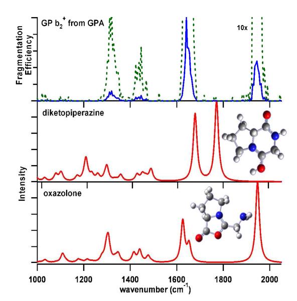
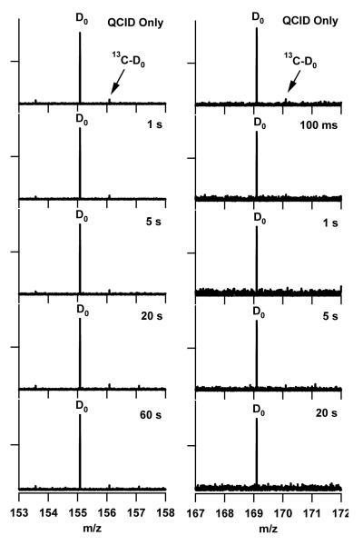
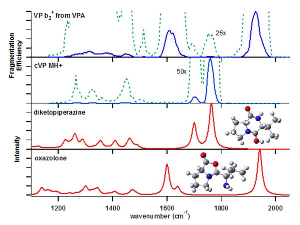
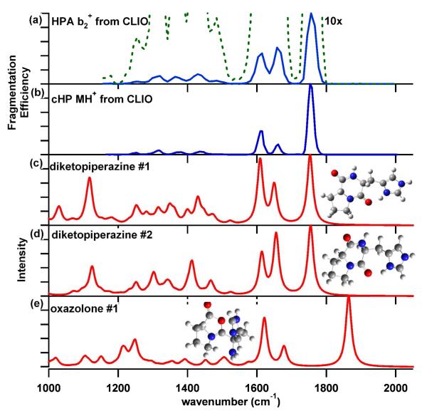
To probe the structural implications of the proline residue on its characteristic peptide fragmentation patterns, in particular its unusual cleavage at its C-terminus in formation of a b(2) ion in XxxProZzz sequences, the structures of a series of proline-containing b(2)(+) ions were studied by using action infrared multiphoton dissociation (IRMPD) spectroscopy and fragment ion hydrogen-deuterium exchange (HDX). Five different Xxx-Pro b(2)(+) ions were studied, with glycine, alanine, isoleucine, valine, or histidine in the N-terminal position. The residues selected feature different sizes, chain lengths, and gas phase basicities to explore whether the structure of the N-terminal residue influences the Xxx-Pro b(2)(+) ion structure. In proteins, the proline side chain-to-backbone attachment causes its peptide bonds to be in the cis conformation more than any other amino acid, although trans is still favored over cis. However, HP is the only b(2)(+) ion studied here that forms the diketopiperazine exclusively. The GP, AP, IP, and VP b(2)(+) ions formed from protonated tripeptide precursors predominantly featured oxazolone structures with small diketopiperazine contributions. In contrast to the b(2)(+) ions generated from tripeptides, synthetic cyclic dipeptides VP and HP were confirmed to have exclusive diketopiperazine structures.


Figures

Figure 1
Figure 1
Action IRMPD spectrum of the GP b2 + ion generated from the protonated tripeptide GPA. Theoretical diketopiperazine (middle) and oxazolone (bottom) spectra, corresponding to the overlaid lowest energy structures calculated at the 6-311++G** level, are shown for comparison. A 10-fold magnified experimental spectrum is shown in dashed green.
Figure 2
Figure 2
HDX behavior of the (left) GP and AP (right) b2 + ions. Exchange time points increase downward, as labeled. The QCID only (no exchange) spectrum is shown at the top to show the presence of the 13C isotopomer of the D0 population and not a D1 population, as confirmed by mass defect analysis.
Figure 3
Figure 3
Action IRMPD spectrum of the AP b2 + ion. Calculated IR spectra from lowest energy diketopiperazine (middle) and oxazolone (bottom) structures are shown for comparison. A 50-fold magnification of the experimental spectrum is shown to highlight the diketopiperazine contribution.
Figure 4
Figure 4
Action IRMPD spectra of the (a) VP b2 + ion generated from the protonated tripeptide VPA and (b) the protonated synthetic diketopiperazine VP (cVP). Magnifications of the experimental spectra are shown in the dashed green line overlaid on each experimental spectrum. Lowest energy (c) diketopiperazine and (d) oxazolone structures are shown for comparison.
Figure 5
Figure 5
HDX behavior of the VP b2 + ion (left), protonated cyclic diketopiperazine VP (middle), and IP b2 + ion (left). Exchange time increases from top to bottom of the figure.
Figure 6
Figure 6
Action IRMPD spectra of the (a) b2 + ion from protonated HPA and (b) protonated diketopiperazine HP. The two calculated lowest-energy diketopiperazine (c) & (d) and oxazolone (e) spectra, along with their corresponding structures, are shown for comparison.
Figure 7
Figure 7
Fragment Ion HDX of the b2 + ions from protonated HPA (left) and cyclo-HP (right). Exchange time increases from top to bottom panel.

Similar articles

Cited by

References

    1. Aebersold R, Mann M. Nature. 2003;422:198–207. - PubMed
    1. Hunt DF, Yates JR, Shabanowitz J, Winston S, Hauer CR. Proc. Nat. Acad. Sci. U. S. A. 1986;83:6233–6237. - PMC - PubMed
    1. Craig R, Beavis RC. Bioinformatics. 2004;20:1466–1467. - PubMed
    1. Elias JE, Gibbons FD, King OD, Roth FP, Gygi SP. Nat. Biotechnol. 2004;22:214–219. - PubMed
    1. Eng JK, McCormack AL, Yates JR. J. Am. Soc. Mass Spectrom. 1994;5:976–989. - PubMed

Publication types