

Progression of ulcerative dermatitis lesions in C57BL/6Crl mice and the development of a scoring system for dermatitis lesions
- PMID: 23312087
- PMCID: PMC3447447
Progression of ulcerative dermatitis lesions in C57BL/6Crl mice and the development of a scoring system for dermatitis lesions
Abstract
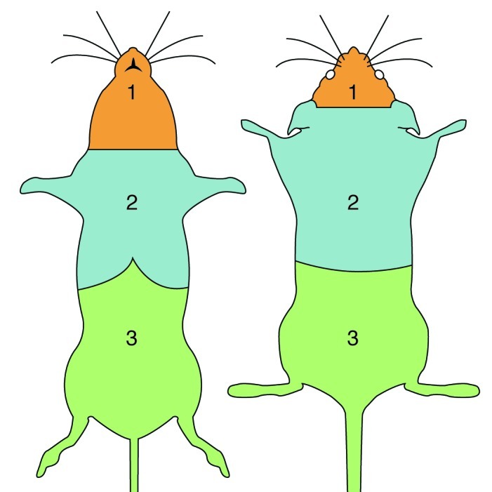
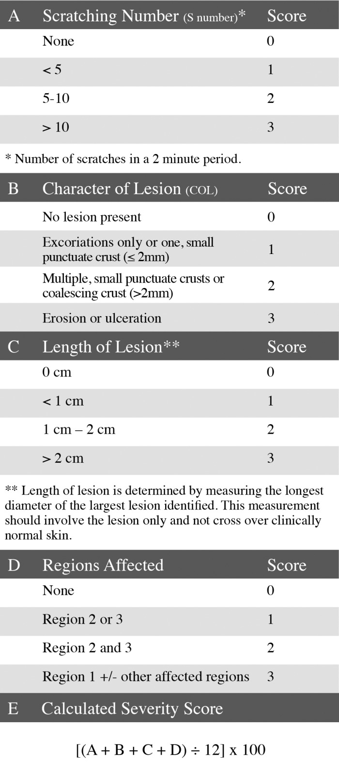
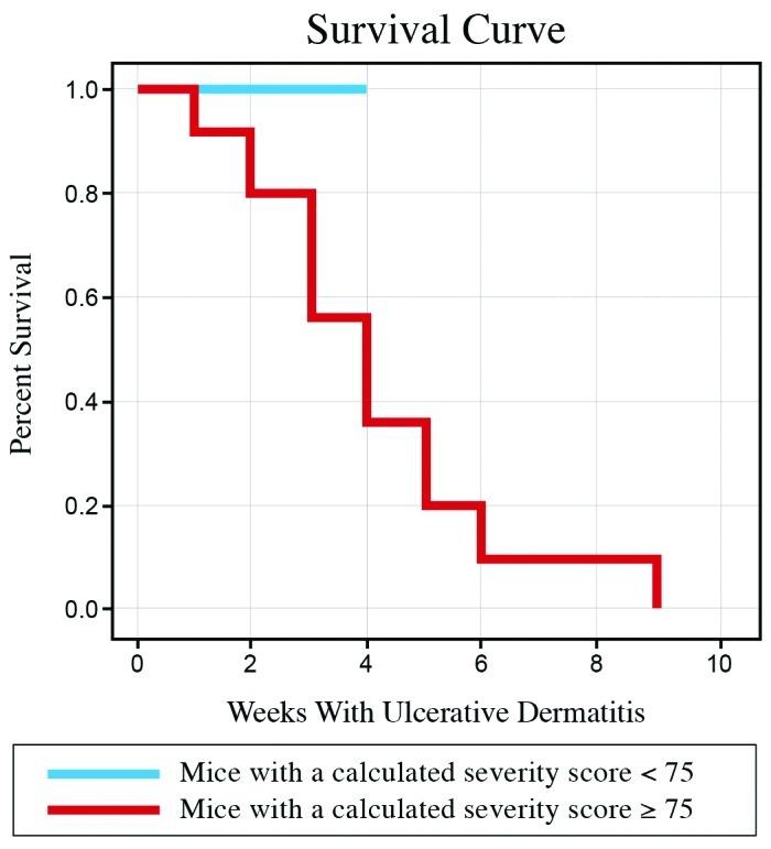
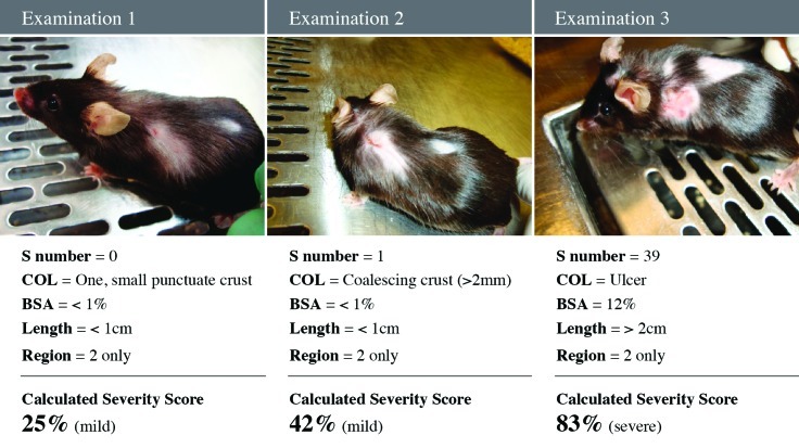
Ulcerative dermatitis (UD) is a common, spontaneous condition in mice with a C57BL/6 background. Although initial lesions may be mild, UD is a progressive disease that often results in ulcerations or debilitating fibrotic contractures. In addition, lesions typically are unresponsive to treatment. Euthanasia is often warranted in severe cases, thereby affecting study outcomes through the loss of research subjects. Because the clinical assessment of UD can be subjective, a quantitative scoring method and documentation of the likely time-frame of progression may be helpful in predicting when animals that develop dermatitis should be removed from a study. Such a system may also be helpful in quantitatively assessing success of various treatment strategies and be valuable to clinical laboratory animal veterinarians. In this 1.5-y, prospective cohort study, we followed 200 mice to monitor the development and course of UD. Mice were examined every 2 wk. A clinical sign (alopecia, pruritus, or peripheral lymphadenopathy) was not identified that predicted development of UD lesions in the subsequent 2-wk period. Once UD developed, pruritus, the character of the lesion (single or multiple crust, coalescing crust, erosion, or ulceration), and the size of the lesion were the only parameters that changed (increased) over the course of the disease. Pruritus was a factor in the rapid progression of UD lesions. We used these findings to develop a quantitative scoring system for the severity of UD. This enhanced understanding of the progression of UD and the quantitative scoring system will enhance the monitoring of UD.
Figures
References
-
- Andrews AG, Dysko RC, Spilman SC, Kunkel RG, Brammer DW, Johnson KJ. 1994. Immune complex vasculitis with secondary ulcerative dermatitis in aged C57BL/6NNia mice. Vet Pathol 31:293–300 - PubMed
-
- Bae CJ, Shim SB, Jee SW, Lee SH, Kim MR, Lee JW, Lee CK, Hwang DY. 2010. IL6, VEGF, KC, and RANTES are a major cause of a high-irritant dermatitis to phthalic anhydride in C57BL/6 inbred mice. Allergol Int 59:389–397 - PubMed
-
- Blackwell BN, Bucci TJ, Hart RW, Turturro A. 1995. Longevity, body weight, and neoplasia in ad-libitum-fed and diet-restricted C57BL6 mice fed NIH31 open-formula diet. Toxicol Pathol 23:570–582 - PubMed
Publication types
MeSH terms
Grants and funding
LinkOut - more resources
Full Text Sources
Other Literature Sources