Review
. 2013 Jan 8;17(1):20-33.
doi: 10.1016/j.cmet.2012.11.012.

Vascular complications of diabetes: mechanisms of injury and protective factors

Affiliations
Review

Vascular complications of diabetes: mechanisms of injury and protective factors

Christian Rask-Madsen et al. Cell Metab. .

Abstract

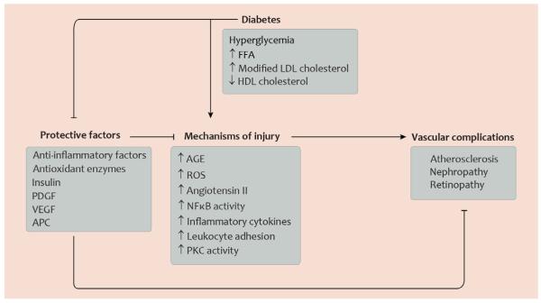
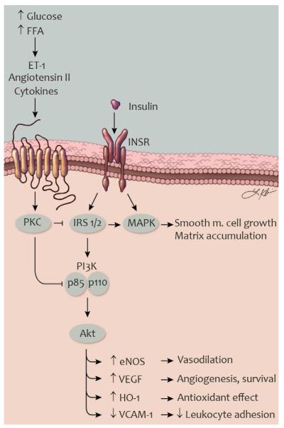
In patients with diabetes, atherosclerosis is the main reason for impaired life expectancy, and diabetic nephropathy and retinopathy are the largest contributors to end-stage renal disease and blindness, respectively. An improved therapeutic approach to combat diabetic vascular complications might include blocking mechanisms of injury as well as promoting protective or regenerating factors, for example by enhancing the action of insulin-regulated genes in endothelial cells, promoting gene programs leading to induction of antioxidant or anti-inflammatory factors, or improving the sensitivity to vascular cell survival factors. Such strategies could help prevent complications despite suboptimal metabolic control.


Figures

Fig. 1
Fig. 1. Selected mechanisms of injury and protective factors determining development of diabetic vascular complications
This diagram illustrates that in the normal state, factors with protective functions in the vasculature can render blood vessels less susceptible to vascular disease and can counteract mechanisms which promote vascular injury. In diabetes, however, glucose and lipid metabolites promote mechanisms of injury and, at the same time, inhibit factors with protective functions in the vasculature. Abbreviations: AGE, advanced glycation end-products; APC, activated protein C; FFA, free fatty acids; HDL, high-density lipoprotein; LDL, low-density lipoprotein; NFκB, nuclear factor κB; PDGF platelet-derived growth factor; PKC, protein kinase C; ROS, reactive oxygen species; VEGF, vascular endothelial growth factor. Artwork by Leah A. Klein.
Fig. 2
Fig. 2. Important histopathological changes during development of atherosclerosis, nephropathy, and retinopathy in diabetes
This schematic illustration shows pathological changes in a coronary artery (left) and in glomerular and retinal capillaries (middle and right, respectively) in diabetes. Some of the main features are accumulation of lipid-laden macrophages in the atherosclerotic plaque and subsequent macrophage apoptosis (left); podocyte apoptosis, thickening of the glomerular basement membrane, and breakdown of the filtration barrier in the renal glomeruli (middle); and pericyte and endothelial cell apoptosis, vascular leakage , and hemorrhage in the retina (right). Proportions, in particular size of the artery relative to the capillaries, are not to scale. Artwork by Leah A. Klein.
Fig. 3
Fig. 3. General abnormalities of vascular function in diabetes
Diagram showing changes in some of the main functions of blood vessels. The diagram should be read like a table, with the affected vessel in each of 3 columns and functions row by row. In arteries, blood flow may be reduced because of atherosclerosi. Glomerular perfusion is increased in early diabetes and the retina becomes ischemic because of insufficient blood flow. Leukocyte adhesion is present in all three vascular beds, but has a particularly important role in atherogenesis. Increased vascular permeability has a prominent role in the glomerular and retinal capillary. Cellular proliferation occurs for vascular smooth muscle cells in the atherosclerotic plaques and for endothelial cells in proliferative diabetic retinopathy. Apoptosis has important implications when it occurs in macrophages in atherosclerosis and is a major characteristic of the histopathology in diabetic nephropathy and retinopathy. Vessels in gray represent a less prominent abnormality for the vessel in question. Artwork by Leah A. Klein.
Fig. 4
Fig. 4. Activation of SHP-1 and inhibition of survival pathways in diabetes
Schematic illustration of mechanisms promoting apoptosis in retinal pericytes and glomerular podocytes. PDGF and VEGF receptor signaling promotes cell survival. In diabetes, activation of SHP-1 can dephosphorylate these receptors and contribute to apoptosis. Abbreviations: MAPK, mitogen-activated protein kinase; NFκB, nuclear factor κB; PDGF, platelet-derived growth factor; PDGFR, PDGF receptor; PI3K, phosphatidylinositol 3-kinase; PKC, protein kinase C; ROS, reactive oxygen species; SHP-1, Src homology-2 domain-containing phosphatase-1; VEGF, vascular endothelial growth factor; VEGFR2, VEGF receptor-2. Artwork by Leah A. Klein.
Fig. 5
Fig. 5. Selective insulin resistance in vascular cells in type 2 diabetes
Schematic illustration of mechanisms causing impaired insulin signaling in vascular endothelial and smooth muscle cells. ET-1, angiotensin II, and other factors can be increased by the metabolic milieu and activate PKC. Activated PKC, in turn, can phosphorylate IRS proteins, the p85 subunit of PI3K, and other signaling molecules. In this state, insulin-stimulated activation of the PI3K signaling is inhibited while signaling through the MAPK pathway is preserved or enhanced. Selective insulin resistance in vascular cells cause impaired vasodilation and angiogenesis, reduced antioxidant effects and increased leukocyte adhesion. Abbreviations (see also fig. 3): eNOS, endothelial nitric oxide synthase; ET-1, endothelin-1; FFA, free fatty acids; HO-1, heme oxygenase-1; INSR, insulin receptor; IRS, insulin receptor substrate; TNF-α, tumor necrosis factor-α; VCAM-1, vascular cell adhesion molecule-1. Artwork by Leah A. Klein.

Similar articles

Cited by

References

    1. Abdouh M, Talbot S, Couture R, Hassessian HM. Retinal plasma extravasation in streptozotocin-diabetic rats mediated by kinin B(1) and B(2) receptors. Br J Pharmacol. 2008;154:136–43. - PMC - PubMed
    1. Aicher A, Heeschen C, Mildner-Rihm C, Urbich C, Ihling C, Technau-Ihling K, Zeiher AM, Dimmeler S. Essential role of endothelial nitric oxide synthase for mobilization of stem and progenitor cells. Nat Med. 2003;9:1370–6. - PubMed
    1. Aiello LP, Bursell SE, Clermont A, Duh E, Ishii H, Takagi C, Mori F, Ciulla TA, Ways K, Jirousek M, Smith LE, King GL. Vascular endothelial growth factor-induced retinal permeability is mediated by protein kinase C in vivo and suppressed by an orally effective beta-isoform-selective inhibitor. Diabetes. 1997;46:1473–80. - PubMed
    1. Aiello LP, Vignati L, Sheetz MJ, Zhi X, Girach A, Davis MD, Wolka AM, Shahri N, Milton RC. Oral protein kinase C beta inhibition using ruboxistaurin: efficacy, safety, and causes of vision loss among 813 patients (1,392 eyes) with diabetic retinopathy in the Protein Kinase C beta Inhibitor-Diabetic Retinopathy Study and the Protein Kinase C beta Inhibitor-Diabetic Retinopathy Study 2. Retina. 2011;31:2084–94. - PubMed
    1. Antonetti DA, Klein R, Gardner TW. Diabetic retinopathy. N Engl J Med. 2012;366:1227–39. - PubMed

Publication types

MeSH terms