. 2013 Jan 8;17(1):35-48.
doi: 10.1016/j.cmet.2012.12.004.

Inhibition of leptin regulation of parasympathetic signaling as a cause of extreme body weight-associated asthma

Affiliations

Inhibition of leptin regulation of parasympathetic signaling as a cause of extreme body weight-associated asthma

Emilio Arteaga-Solis et al. Cell Metab. .

Erratum in

  • Cell Metab. 2013 Mar 5;17(3):463-4

Abstract

Impaired lung function caused by decreased airway diameter (bronchoconstriction) is frequently observed whether body weight is abnormally high or low. That these opposite conditions affect the airways similarly suggests that the regulation of airway diameter and body weight are intertwined. We show here that, independently of its regulation of appetite, melanocortin pathway, or sympathetic tone, leptin is necessary and sufficient to increase airway diameter by signaling through its cognate receptor in cholinergic neurons. The latter decreases parasympathetic signaling through the M(3) muscarinic receptor in airway smooth muscle cells, thereby increasing airway diameter without affecting local inflammation. Accordingly, decreasing parasympathetic tone genetically or pharmacologically corrects bronchoconstriction and normalizes lung function in obese mice regardless of bronchial inflammation. This study reveals an adipocyte-dependent regulation of bronchial diameter whose disruption contributes to the impaired lung function caused by abnormal body weight. These findings may be of use in the management of obesity-associated asthma.


Figures

Figure 1
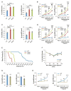
Figure 1. High and Low Fat Mass Results in Bronchoconstriction
(A) Diet-induced obese (DIO) WT mice gain significant weight after 12 weeks on a high-fat diet. (B) Total respiratory resistance (R) and airway resistance (Rn) are increased in DIO-WT mice. (C) DIO-WT mice have innate airway hyperreactivity (AHR) to methacholine. (D) Endurance is severely limited in DIO-WT mice when placed on a treadmill. (E and F) Lypodystrophic, “fat-free,” mice have increased baseline R, Rn, and AHR. For all panels, n = 10 per group. *p < 0.05, **p < 0.01, and ***p < 0.001. Error bars represent SEM. See also Figure S1.
Figure 2
Figure 2. Leptin Regulates Bronchial Diameter Independently of Appetite and Body Weight
(A and B) Severely obese 8-week-old ob/ob and db/db mice demonstrated increased R, Rn, and AHR (n = 10 per group, *db/db versus WT, #ob/ob versus WT). (C and D) Moderately overweight 5-week-old ob/ob and db/db mice demonstrated increased R, Rn, and AHR (n = 12 per group, *db/db versus WT, #ob/ob versus WT). (E) Endurance is decreased in 5-week-old ob/ob and db/db compared to WT mice (n = 8 per group). (F) l/l mice have normal insulin sensitivity (n = 5 per group). (G and H) Increased leptin signaling in l/l mice leads to a decrease in baseline R, Rn, and AHR compared to WT controls (n = 8 per group). For all panels, */#p < 0.05, **/##p < 0.01, and ***/###p < 0.001. Error bars represent SEM. See also Figure S2.
Figure 3
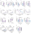
Figure 3. Leptin Regulates Bronchial Diameter via a Central Pathway
(A) Mice with deletion of ObRb in all neurons (ObRbneuron-ERT2/) have normal insulin sensitivity (n = 5 per group). (B and C) ObRbneuron-ERT2/ mice have decreased R, Rn, and AHR compared to tamoxifen-treated Nestincre-ERT2 and ObRbfl/fl controls (n = 8 per group, *ObRbneuron-ERT2/ versus Nestincre-ERT2). (D–F) Leptin (Lep) ICV infusion (4 ng/hr for 5 days) improves baseline R, Rn, and AHR, and endurance in ob/ob, not in db/db mice (n = 8 per group, *ob/ob versus WT, #ob/ob versus ob/ob-Lep). (G) ObRb localizes in cholinergic-containing nuclei including the dorsal motor vagus (DMVN) (highlighted area) nucleus and facial motor nucleus (Mo7) (arrows) and is absent in mice with an inducible deletion of ObRb in Ach-producing neurons (ObRbAche-ERT2/). (H–J) ObRbAche-ERT2/ mice have normal insulin sensitivity and increased baseline R, Rn, and AHR compared to tamoxifen-treated Achecre-ERT2 and ObRbfl/fl controls (n = 10 per group). *ObRbAche-ERT2/ versus Achecre-ERT2. For all panels, */#p < 0.05, **/##p < 0.01, and ***/###p < 0.001. Error bars represent SEM. See also Figure S3.
Figure 4
Figure 4. Leptin Regulates Bronchial Diameter by Inhibiting Parasympathetic Nervous System Signaling in ASM Cells through the M3R
(A) Autonomic nervous system blockade by hexamethonium causes a greater decrease in Rn in ob/ob than in WT mice (n = 6 per group, arrow indicates time of hexamethonium administration). (B) Bilateral cervical vagotomy causes a greater decrease in Rn in ob/ob than in WT mice (n = 6 per group, arrow indicates time of vagotomy). (C) Elevated acetylcholine (Ach) levels in ob/ob brain and lungs are decreased by leptin (Lep) ICV infusion (4 ng/hr for 5 days) (n = 5 per group). (D) Decreased cholinesterase activity in ob/ob brain and lungs is improved with leptin (Lep) ICV (n = 5 per group). (E) M2R and M3R muscarinic receptors are the highest expressed cholinergic receptors in the mouse lung. (F) M2R/ mice have increased R and Rn compared to WT controls (n = 6 per group). (G) Global M3R/ and smooth muscle-specific inducible deletion of M3R (M3RSM-ERT2/) decrease baseline R and Rn in mice compared to WT and tamoxifen-treated SMMHC-CreERT2 control mice, respectively (n = 8 per group). (H and I) M3RSM-ERT2/ mice have normal insulin sensitivity and weight compared to tamoxifen-treated SMMHC-CreERT2 control mice (n = 5 per group). (J) Primary M3RSM-ERT2/ ASM cells have a marked reduction in M3R expression levels compared to control ASM cells, but not in the brainstem (n = 6 per group). (K) M3RSM-ERT2/ mice have no response to methacholine compared to tamoxifen-treated SMMHC-CreERT2 and WT littermate controls, confirming M3R deletion in ASM cells (n = 8 per group, *WT versus M3RSM-ERT2/, #SMMHC-CreERT2 versus M3RSM-ERT2/). (L and M) Removing one allele of M3R in smooth muscle of ob/ob mice normalizes their baseline R, Rn, and AHR, as leptin ICV does (n = 8 per group, *ob/ob versusob/ob; M3RSM-ERT2/). For all panels, */#p < 0.05, **/##p < 0.01, and ***/###p < 0.001. Error bars represent SEM. See also Figure S4.
Figure 5
Figure 5. Central Leptin-Dependent Regulation of Bronchial Diameter in Diet-Induced Obese WT Mice
(A–D) Diet-induced obese (DIO)-WT mice have increased levels of circulating leptin, food intake, and UCP-1 expression in brown fat and levels of urine norepinephrine, all suggestive of a state of leptin resistance (n = 8 per group). (E and F) Increased baseline R, Rn, and AHR in DIO-WT mice reversed by leptin (Lep) ICV infusion (12 ng/hr for 5 days) (n = 8 per group, *DIO-WT versus DIO-WT Lep). (G) Decreased endurance in DIO-WT mice reversed by leptin (Lep) ICV (12 ng/hr for 5 days) or inducible deletion of one allele of M3R in smooth muscle cells (M3RSM-ERT2+/) (n = 8 per group). (H) Bilateral cervical vagotomy causes a greater decrease in Rn in DIO-WT than in WT mice (n = 8 per group, arrow indicates time of vagotomy). (I) High acetylcholine (Ach) levels in brain and lungs of DIO-WT mice corrected by leptin (Lep) ICV (12 ng/hr for 5 days) infusion (n = 6 per group). (J) Decreased cholinesterase activity in DIO-WT improved by leptin ICV (n = 6 per group). (K–M) Obese DIO-M3R+/ and DIO-M3RSM-ERT2+/ mice have baseline R, Rn, and AHR comparable to lean tamoxifen-treated SMCre-ERT2 and WT mice (n = 8 per group, *DIO-WT versus DIO-M3R+/, #DIO-WT versus DIO-M3RSM-ERT2+/). For all panels, */#p < 0.05, **/##p < 0.01, and ***/###p < 0.001. Error bars represent the SEM. See also Figure S4.
Figure 6
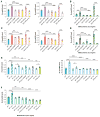
Figure 6. Leptin Regulation of Bronchial Diameter Occurs Independently of Local Inflammation
(A) Bronchoalveolar lavage analysis demonstrates no difference in inflammatory cells in DIO-WT, ob/ob, “fat-free,” or db/db compared to the ovalbumin (ova)-sensitized and challenged mice (n = 8 per group). (B) Periodic acid-Shiff staining fails to detect goblet cell (arrows) hyperplasia in DIO-WT, ob/ob, ‘fat-free,” or db/db mice compared to ova mice. (C–F) Leptin (Lep) ICV infusion in WTova and ob/obova mice decreased Rn and AHR compared to control (n = 12 per group, D-*WTova versus WTova Lep, F-*ob/obova versus ob/obova Lep). (G) Leptin ICV infusion failed to change the levels of inflammatory cells in WTova, DIO-WTova, ob/obova mice (n = 8 per group). (H) Leptin ICV infusion failed to change the goblet cell (arrows) hyperplasia in WTova, DIO-WTova, ob/obova mice. For all panels, *p < 0.05, **p < 0.01, and ***p < 0.001. Error bars represent the SEM. See also Figure S5.
Figure 7
Figure 7. Single Anticholinergic Agent Improves Airway Resistance in Obese Mice
(A, C, and E) Nebulized tiotropium (Tio) (200 μl of 50 μg/ml solution once a day for 2 days), 4-diphenylacetoxy-N-methylpiperidine methiodide (4-DAMP) (200 μl of 100 μg/ml solution 30 min prior to airway measurements), and darifenacin (Dari) (200 μl of 100 μg/ml solution 30 min prior to airway measurements) therapies normalize R and Rn in DIO-WT, DIO-WTova, ob/ob, ob/obova, and WTova male mice (n = 6 per group). (B, D, and F) Anticholinergic therapy blocked the response to nebulized methacholine (20 μl of 12.5 mg/ml solution) in DIO-WT, DIO-WTova, ob/ob, ob/obova, and WTova-treated compared to control mice, indicating proper therapeutic drug delivery (n = 6 per group). For all panels, *p < 0.05, **p < 0.01, and ***p < 0.001. Error bars represent the SEM.

Similar articles

Cited by

References

    1. Barnes PJ. The pharmacological properties of tiotropium. Chest. 2000;117(2, Suppl):63S–66S. - PubMed
    1. Björnholm M, Münzberg H, Leshan RL, Villanueva EC, Bates SH, Louis GW, Jones JC, Ishida-Takahashi R, Bjørbaek C, Myers MG., Jr Mice lacking inhibitory leptin receptor signals are lean with normal endocrine function. J Clin Invest. 2007;117:1354–1360. - PMC - PubMed
    1. Butler AA, Kesterson RA, Khong K, Cullen MJ, Pelleymounter MA, Dekoning J, Baetscher M, Cone RD. A unique metabolic syndrome causes obesity in the melanocortin-3 receptor-deficient mouse. Endocrinology. 2000;141:3518–3521. - PubMed
    1. Callaerts-Vegh Z, Evans KL, Dudekula N, Cuba D, Knoll BJ, Callaerts PF, Giles H, Shardonofsky FR, Bond RA. Effects of acute and chronic administration of beta-adrenoceptor ligands on airway function in a murine model of asthma. Proc Natl Acad Sci USA. 2004;101:4948–4953. - PMC - PubMed
    1. Canning BJ. Reflex regulation of airway smooth muscle tone. J Appl Physiol. 2006;101:971–985. - PubMed

Publication types

MeSH terms