Review
. 2013 Apr;36(4):237-47.
doi: 10.1016/j.tins.2012.12.002. Epub 2013 Jan 8.

Modeling cognitive dysfunction in neurofibromatosis-1

Affiliations
Review

Modeling cognitive dysfunction in neurofibromatosis-1

Kelly A Diggs-Andrews et al. Trends Neurosci. 2013 Apr.

Abstract

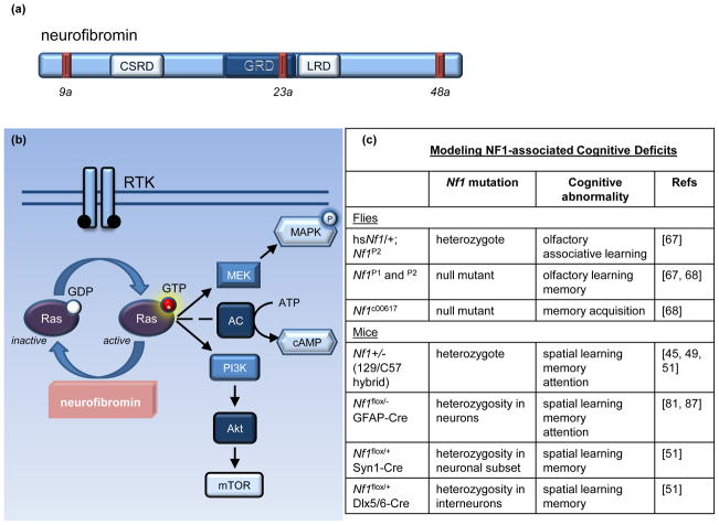
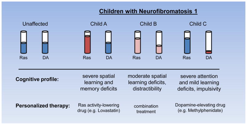
Cognitive dysfunction, including significant impairments in learning, behavior, and attention, is found in over 10% of children in the general population. However, in the common inherited cancer predisposition syndrome, neurofibromatosis type 1 (NF1), the prevalence of these cognitive deficits approaches 70%. As a monogenic disorder, NF1 provides a unique genetic tool to identify and dissect mechanistically the molecular and cellular bases underlying cognitive dysfunction. In this review, we discuss Nf1 fly and mouse systems that mimic many of the cognitive abnormalities seen in children with NF1. Further, we describe discoveries from these models that have uncovered defects in the regulation of Ras activity, cAMP generation, and dopamine homeostasis as key mechanisms important for cognitive dysfunction in children with NF1.


Figures

Figure 1
Figure 1. Neurofibromin structure and function
(a) Neurofibromin is a 2818 amino acid protein that contains multiple alternatively spliced exons (9a, 23a, and 48a shown as red bars) and encodes several distinct functional domains, including a cysteine-rich domain (CSRD), a Ras-GAP domain (GRD), and a leucine repeat domain (LRD) [, –30]. (b) Neurofibromin serves as a negative regulator of Ras by accelerating the hydrolysis of the GTP-bound active Ras, producing inactive GDP-bound Ras [–34]. Upon receptor tyrosine kinase (RTK) activation, Ras guanosine exchange (GDP to GTP) promotes Ras activity. Activated Ras, in turn, stimulates its downstream effectors, including MEK/MAPK and PI3K/Akt/mTOR. Ras can also positively regulate adenylate cyclase (AC) activity in some cell types. (c) Current fly and mouse models of NF1-associated learning and attention abnormalities. Abbreviations: GFAP, glial fibrillary acidic protein; Syn1, synapsin 1; Dlx5/6, distal-less homeobox 5 and 6.
Figure 2
Figure 2. Neurofibromin regulation of neuronal morphology is cAMP-mediated
(a) Neuronal morphology (growth cone areas and neurite length) is attenuated by reduced neurofibromin expression and cAMP levels. Top panel: Primary cultured Nf1+/− hippocampal neurons have decreased neurite lengths (not shown) and growth cone areas as a result of reduced cAMP generation [73]. Middle panel: Stimulating adenylyl cyclase (AC) with Forskolin (FOR) elevates cAMP levels and rescues growth cone areas in Nf1+/− hippocampal neurons. Bottom panel: Decreasing cAMP levels in wild-type neurons by blocking adenylyl cyclase activity with the AC inhibitor 2,3-dideoxyadenosine (DDA) blunts growth cone areas, comparably to Nf1+/− neurons. Scale bar = 20μm. Adapted, with permission, from [73]. (b) Schematic diagram illustrating mechanism of cAMP-mediated morphological changes. In neurons, low cAMP generation due to reduced Nf1 expression, in turn, leads to decreased PKA activation, RhoA/ROCK activity, and MLC phosphorylation. These changes contribute to impaired actin cytoskeletal dynamics, resulting in smaller growth cones and shortened axons [74]. Abbreviations: GPCR, G protein-coupled receptor; MLC, myosin light chain; PKA, protein kinase A; ROCK, Rho-associated protein kinase.
Figure 3
Figure 3. Role of dopamine in NF1-associated behavior
(a) Tyrosine hydroxylase (TH)-positive neurons in the substantia nigra project to the striatum and modulate dopamine signaling. Presynaptically, TH converts tyrosine to L-DOPA. Upon release, dopamine returns to the presynaptic dopamine pool by binding dopamine transporters or it activates adenylyl cyclase (AC) by binding post-synaptic dopamine receptors. AC activation stimulates cAMP generation and dopamine- and cAMP-regulated phosphoprotein-32 (DARPP32) phosphorylation in the striatum. (b) In Nf1 mutant mice, TH expression is reduced, leading to lower striatal dopamine levels and lower effector signaling, as indicated by reduced DARPP32 phosphorylation [81]. Abnormal dopamine homeostasis also results in attention system dysfunction in the Nf1 mutant model [75, 81]. (c) Treatment with drugs that increase dopamine synthesis (L-DOPA), exogenous dopamine levels (dopamine), or block dopamine reuptake [eg. methylphenidate (MPH)] restores striatal dopamine levels and reverses the attention phenotype in Nf1 mutant mice [75, 81]. Abbreviations: GPCR, G-protein coupled receptors.
Figure 4
Figure 4. Factors Influencing the NF1 Cognitive Phenotype
The specific cognitive phenotype observed in any given individual with NF1 reflects the confluence of genomic, molecular, cellular, and environmental factors. The specific germline NF1 gene mutation, allele expression, and genomic modifying events (including methylation), may influence clinical heterogeneity and create variations in neurofibromin expression in different cell types. Heterogeneity in neuronal subpopulations may also factor into the overall cognitive phenotype. In this manner, the relative contributions of defects in different populations of CNS neurons (e.g., GABA-ergic, dopaminergic) may lead to a distinct spectrum of cognitive and behavioral abnormalities. Similarly, less well-studied characteristics, such as patient sex, age, and NF1-associated brain abnormalities (e.g., T2-hyperintensities), may also contribute to the observed patient cognitive profile. In addition, the effects of abnormal signaling through specific molecular pathways (ie. signaling heterogeneity) could likewise differentially impact distinct neuronal populations and lead to unique cognitive phenotypes. While understudied to date, it is also possible that NF1+/− oligodendrocytes, microglia, and/or astrocytes contribute to abnormal neuronal function as a result of disrupted axonal signal propagation, impaired synaptic pruning, and/or changes in glutamate or other neurotransmitter availability.
Figure 5
Figure 5. Model of clinical heterogeneity and implications for treatment
The cognitive profile of children with NF1 likely includes a diverse set of learning, memory, attention, and motor deficits, each linked to a distinct molecular abnormality (i.e. increased Ras, reduced cAMP, or low dopamine). In this regard, each child with NF1 has a different level of Ras, dopamine, and cAMP (not shown) signaling that collectively contribute to the overall cognitive phenotype. Identifying the primary set of cellular and molecular abnormalities may lead to individualized treatments with a higher likelihood of improving the neurocognitive deficits specific to that child or adult with NF1.

References

    1. Hyman SL, et al. The nature and frequency of cognitive deficits in children with neurofibromatosis type 1. Neurology. 2005;65:1037–1044. - PubMed
    1. Mautner VF, et al. Treatment of ADHD in neurofibromatosis type 1. Developmental medicine and child neurology. 2002;44:164–170. - PubMed
    1. Hyman SL, et al. Learning disabilities in children with neurofibromatosis type 1: subtypes, cognitive profile, and attention-deficit-hyperactivity disorder. Developmental medicine and child neurology. 2006;48:973–977. - PubMed
    1. Ferner RE. The neurofibromatoses. Practical neurology. 2010;10:82–93. - PubMed
    1. Wallace MR, et al. Type 1 neurofibromatosis gene: identification of a large transcript disrupted in three NF1 patients. Science. 1990;249:181–186. - PubMed

Publication types