. 2013 Jan 9;77(1):58-69.
doi: 10.1016/j.neuron.2012.10.032.

Notch inhibition induces cochlear hair cell regeneration and recovery of hearing after acoustic trauma

Affiliations

Notch inhibition induces cochlear hair cell regeneration and recovery of hearing after acoustic trauma

Kunio Mizutari et al. Neuron. .

Erratum in

  • Neuron. 2013 Apr 24;78(2):403
  • Neuron. 2015 Apr 8;96(1):341

Abstract

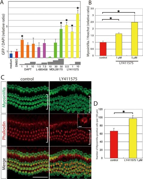
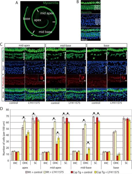
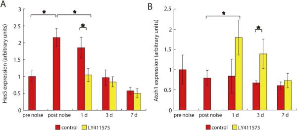
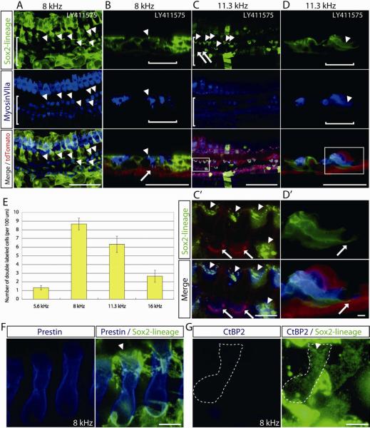
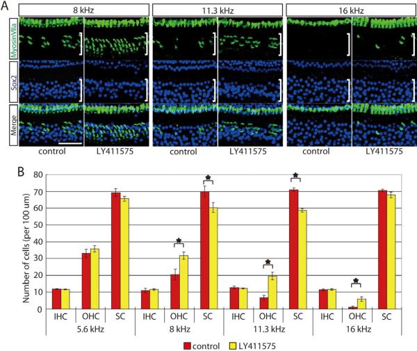
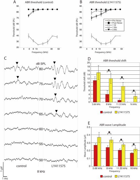
Hearing loss due to damage to auditory hair cells is normally irreversible because mammalian hair cells do not regenerate. Here, we show that new hair cells can be induced and can cause partial recovery of hearing in ears damaged by noise trauma, when Notch signaling is inhibited by a γ-secretase inhibitor selected for potency in stimulating hair cell differentiation from inner ear stem cells in vitro. Hair cell generation resulted from an increase in the level of bHLH transcription factor Atoh1 in response to inhibition of Notch signaling. In vivo prospective labeling of Sox2-expressing cells with a Cre-lox system unambiguously demonstrated that hair cell generation resulted from transdifferentiation of supporting cells. Manipulating cell fate of cochlear sensory cells in vivo by pharmacological inhibition of Notch signaling is thus a potential therapeutic approach to the treatment of deafness.


Figures

Figure 1
Figure 1. In vitro activity of γ-secretase inhibitors in hair cell induction
(A) Relative ratio of nGFP-positive cells to DAPI-positive cells after treatment of inner ear spheres made from Math1-nGFP mice with γ-secretase inhibitors at the indicated concentrations (μM) reveals that LY411575 had the greatest potency of 4 inhibitors tested for hair cell induction. Data were normalized to control values obtained by addition of DMSO. Asterisks indicate p < 0.01. (B) Ratio of myosin VIIa (labels hair cells) to Hoechst-positive cells induced by LY411575 was calculated relative to DMSO-treated spheres from organ of Corti. (C) Explant cultures of the organ of Corti from P1 mice cultured for 72 h in the presence of DMSO or LY411575 (1 μM) had ectopic hair cells (myosin VIIa; green) in the outer hair cell region (white bracket). Ectopic hair cells were positive for phalloidin (labels the hair bundle and cuticular plate; shown in red). Inset is a high-power view (scale bar is 2 μM) of a phalloidin-stained hair cell showing bundle structure. (D) An increase in myosin VIIa-positive cells per 100 μm of the cultured organ of Corti explants from P1 mice was found 72 h after LY411575 treatment. In all graphs, error bars show the standard error of the mean. Scale bar is 50 μm.
Figure 2
Figure 2. Hair cell replacement after LY411575 treatment of organ of Corti explants from mice subjected to ablation of hair cells
(A) Hair cells can be seen throughout the neonatal organ of Corti in a whole mount labeled for myosin VIIa. (B) Three rows of outer (white bracket; OHC1 – 3) and one row of inner hair cells (IHC) can be seen in a P3 organ of Corti explant after staining for myosin VIIa. Deiters' cells (DC1 - 3) and Hensen cells (HC) in the outer hair cell region are positive for Sox2. (C) Organ of Corti explants from Pou4f3-Cre; Mos-iCsp3 double-transgenic mice subjected to dimerizer-induced hair cell ablation and cultured for 3 d in the presence of LY411575 had an increased number of myosin VIIa-positive cells in the outer hair cell region (white bracket) compared to the carrier-treated explant. The same region had a decreased number of Sox2-positive cells relative to the control. A high power view (scale bar, 2 μM) of phalloidin-stained tissue shows the hair cell stereociliary bundles (inset). (D) The number of outer hair cells at the mid-apex and mid-base was increased in the LY411575 treated as compared to the control cochleae in the hair cell-ablated samples (Csp Tg). Increased numbers of hair cells were also seen after LY411575 treatment of wild-type organ of Corti (Wt) at the apex, mid-apex and mid-base. In both cases the increase in the number of hair cells was accompanied by a decrease in the number of supporting cells. The error bars are standard error of the mean (n = 7 in each group). Asterisks indicate p < 0.05. All scale bars are 50 μm.
Figure 3
Figure 3. Time course of Hes5 and Atoh1 mRNA expression in the cochlea with or without LY411575 after noise exposure
(A) Elevated levels of Hes5 after noise exposure were diminished in response to LY411575 treatment and reached the pre noise level. Without inhibitor, expression levels of Hes5 in the cochlea increased 1 d after noise exposure and remained elevated compared to the pre noise level for up to 2 d. Samples for qRT-PCR were taken before exposure to noise (pre noise), at the time (day 0) of drug treatment (post noise), at day 1 of drug treatment (d 1), day 3 of drug treatment (d 3), and day 7 of drug treatment (d 7). mRNA expression levels were calculated relative to the pre-noise level. (B) Treatment with LY411575 significantly increased the expression of Atoh1 compared to the opposite, untreated ear 1 d after noise exposure. Increased levels were detected 1 d after drug treatment (d 1) and remained elevated 3 d after drug treatment (d 3; n = 9 in each group. Error bars are standard error of the mean. Asterisks indicate p < 0.05.
Figure 4
Figure 4. Lineage tracing of supporting cells in noise-exposed cochleae treated in vivo with a γ-secretase inhibitor
(A) Double-labeled cells (arrowheads) positive for Sox2 lineage (GFP) and myosin VIIa (blue) were observed in the outer hair cell area (white bracket) in cochlear tissues from deafened mice carrying the Sox2-CreER as well as the Cre reporter transgene, mT/mG, 1 month after LY411575 treatment. Hair cell co-labeling with the lineage tag indicates derivation from a Sox2-positive cell and is thus evidence for regenerated hair cells after deafening in the mature mouse cochlea by transdifferentiation of supporting cells. These confocal xy-projection images of LY411575-treated ears from Sox2-CreER; mT/mG double transgenic mice are in the 8 kHz area of the cochlear longitudinal frequency map. (B) Confocal xz-projections from the same area as A show that myosin VIIa-positive cells in the medial part of the outer hair cell area (white bracket) had GFP-positive hair bundle structures, indicating a Sox2 lineage (arrowhead). The cell shown was attached to the basement membrane (arrow) similar to a supporting cell. (C) Cells double-labeled for myosin VIIa (blue) and Sox2 lineage (green) were observed (arrowheads) in the outer hair cell area (white bracket) in the 11.3 kHz region in this xy projection from a deafened cochlea 1 month after LY411575 treatment. Original hair cells have red hair bundles and new, Sox2-lineage hair cells have green (GFP-positive) bundles. (C') High power view of hair cells with their original (red) bundles (arrows) adjacent to cells with new (green) bundles (arrowheads) derived from Sox2-positive cells. (D) Cross section from the same area as C shows that myosin VIIa, Sox2-lineage double-labeled cells in the outer hair cell area (white bracket) spanned the epithelium from the basement membrane to the endolymphatic surface. (D') The cell shown is attached to the basement membrane (arrow) and its nucleus is at the base of the cell. (E) Quantification of the GFP (Sox2 lineage) and myosin VIIa double-labeled cells in the outer hair cell region 1 month after treatment with LY411575 in deafened mice at frequency-specific cochlear areas (n = 5 in each group). Error bars are standard error of mean. (F) Cells double-labeled for prestin (blue) and Sox2 lineage (green) were observed in the 8 kHz region in this xy projection from a deafened cochlea 1 month after LY411575 treatment. Sox2-lineage hair cell has green (GFP-positive) bundles (white arrowhead). (G) Sox2-lineage hair cells (white broken line) were negative for CtBP2, which labels inner hair cell synaptic ribbons. White arrowhead indicates hair cell bundle. A–D: Scale bars are 50 μm. F, G: Scale bars are 5 μm.
Figure 5
Figure 5. Hair cells in damaged mature cochlea treated with LY411575 in vivo
(A) The number of hair cells (green; myosin VIIa) in the outer hair cell region (white brackets) of deafened cochleae at 8, 11.3, and 16 kHz areas was increased compared to the control ear (right ear treated with carrier) 3 months after treatment with LY411575 (left ear), and the increase was accompanied by a decrease in the number of supporting cells (blue: Sox2) in the same regions in these whole mount confocal xy-projections. (B) Significant differences in the numbers of hair cells and supporting cells were observed in the outer hair cell area at 8 and 11.3 kHz regions of treated (left) ears 3 months after treatment with LY411575 as compared to the values in the contralateral carrier-treated ear of deafened mice (n = 5 in each group). All scale bars are 50 μm. Error bars are standard error of the mean and asterisks indicate p < 0.05.
Figure 6
Figure 6. Measurement of ABR in deafened ears after LY411575 treatment
(A, B) A decrease in ABR thresholds at low frequencies (up to 16 kHz) in the left, LY411575-treated ear (B) compared to the right, control ear (A) was apparent in ABR thresholds in recordings made at 7 frequencies from 5.66 to 45.25 kHz with the following time course. Before noise exposure (Pre Noise: open circles), 1 d after noise exposure (Post Noise: filled circles), 1 week after drug treatment (1 W: open squares), 1 month after treatment (1 Mo: crosses), and 3 months after treatment (3 Mo: filled triangles) (n = 5 in each group). When no response was observed at 80 dB (maximum acoustic output of the system) the threshold was designated as 85 dB. (C) An example of 8 kHz ABR waves recorded 3 months after drug treatment from the same mouse. Arrowheads show the peaks with the largest peak-to-peak amplitude. In the LY411575-treated ear, the peak could first be detected at 65 dB, while on the control side the peak could first be detected at 75 dB. (D, E) The differences in threshold (D) and wave I amplitude (E) 3 months after drug treatment compared to 1 d after noise exposure between control and LY411575-treated ears at 8, 11.33, and 16 kHz (asterisks) were significant (n = 5 in each group). Error bars are standard error of the mean.

Comment in

References

    1. Adam J, Myat A, Le Roux I, Eddison M, Henrique D, Ish-Horowicz D, Lewis J. Cell fate choices and the expression of Notch, Delta and Serrate homologues in the chick inner ear: parallels with Drosophila sense-organ development. Development. 1998;125:4645–4654. - PubMed
    1. Arnold K, Sarkar A, Yram MA, Polo JM, Bronson R, Sengupta S, Seandel M, Geijsen N, Hochedlinger K. Sox2(+) adult stem and progenitor cells are important for tissue regeneration and survival of mice. Cell Stem Cell. 2011;9:317–329. - PMC - PubMed
    1. Batts SA, Shoemaker CR, Raphael Y. Notch signaling and Hes labeling in the normal and drug-damaged organ of Corti. Hear Res. 2009;249:15–22. - PMC - PubMed
    1. Cafaro J, Lee GS, Stone JS. Atoh1 expression defines activated progenitors and differentiating hair cells during avian hair cell regeneration. Dev Dyn. 2007;236:156–170. - PubMed
    1. Caiazzo M, Dell'Anno MT, Dvoretskova E, Lazarevic D, Taverna S, Leo D, Sotnikova TD, Menegon A, Roncaglia P, Colciago G, et al. Direct generation of functional dopaminergic neurons from mouse and human fibroblasts. Nature. 2011;476:224–227. - PubMed

Publication types

MeSH terms

Substances