. 2013 Sep 1;74(5):357-66.
doi: 10.1016/j.biopsych.2012.12.003. Epub 2013 Jan 8.

Mifepristone alters amyloid precursor protein processing to preclude amyloid beta and also reduces tau pathology

Affiliations

Mifepristone alters amyloid precursor protein processing to preclude amyloid beta and also reduces tau pathology

David Baglietto-Vargas et al. Biol Psychiatry. .

Abstract

Background: Increased circulating glucocorticoids are features of both aging and Alzheimer's disease (AD), and increased glucocorticoids accelerate the accumulation of AD pathologies. Here, we analyzed the effects of the glucocorticoid receptor antagonist mifepristone (RU486) in the 3xTg-AD mouse model at an age where hippocampal damage leads to high circulating corticosterone levels.

Methods: The effects of mifepristone were investigated in 3xTg-AD mice using a combination of biochemical, histological, and behavior analyses.

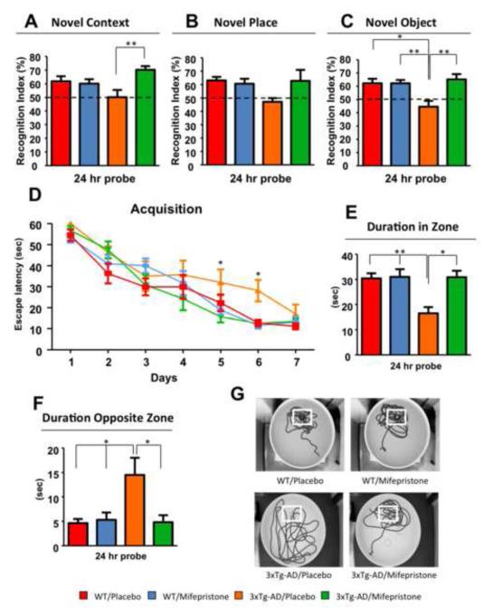
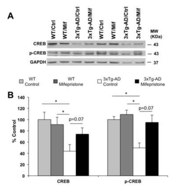
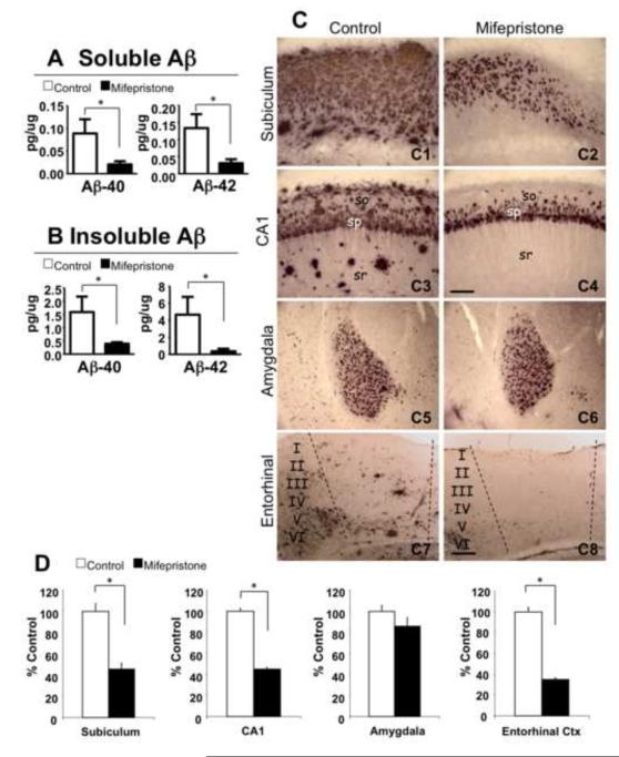
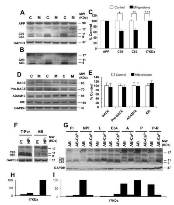
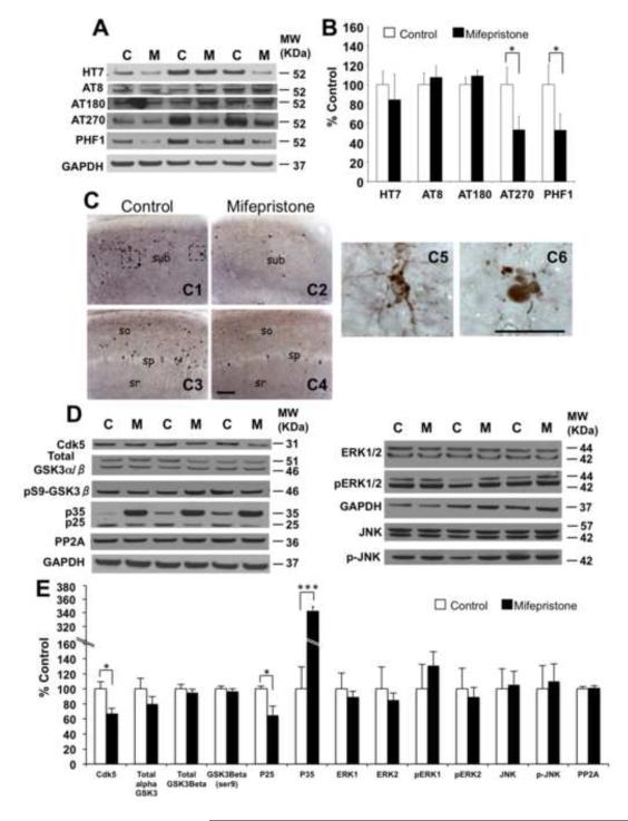
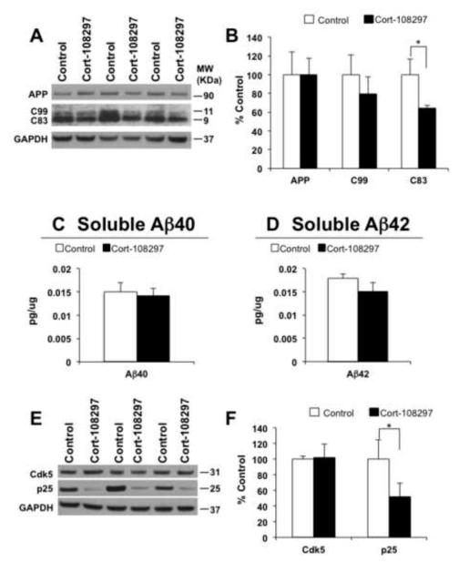
Results: Mifepristone treatment rescues the pathologically induced cognitive impairments and markedly reduces amyloid beta (Aβ)-load and levels, as well as tau pathologies. Analysis of amyloid precursor protein (APP) processing revealed concomitant decreases in both APP C-terminal fragments C99 and C83 and the appearance of a larger 17-kDa C-terminal fragment. Hence, mifepristone induces a novel C-terminal cleavage of APP that prevents it being cleaved by α- or β-secretase, thereby precluding Aβ generation in the central nervous system; this cleavage and the production of the 17-kDa APP fragment was generated by a calcium-dependent cysteine protease. In addition, mifepristone treatment also reduced the phosphorylation and accumulation of tau, concomitant with reductions in p25. Notably, deficits in cyclic-AMP response element-binding protein signaling were restored with the treatment.

Conclusions: These preclinical results point to a potential therapeutic role for mifepristone as an effective treatment for AD and further highlight the impact the glucocorticoid system has as a regulator of Aβ generation.

Keywords: 3xTg-AD; Alzheimer's disease; Amyloid beta; glucocorticoids; mifepristone; tau.


Figures

Figure 1
Figure 1. Mifepristone rescues cognitive impairments in 3xTg-AD mice
A-C) Significant improvements in memory by novel context (A) and object (C) recognition were observed between mifepristone and vehicle treated 3xTg-AD mice. B) No significant differences were detected in novel place. D) Mice were trained on the spatial reference version of the Morris water maze (MWM; N=8-10 per group). Acquisition curves shown for the 7 days of training on the MWM. Mifepristone treatment reduces spatial memory deficits during training. E-F) Mifepristone-treated mice shown significant improvements in duration spent in the target zone (E) and opposite zone (F). G) Representative path tracings of the probe test session (white box represent localization of platform target). Ntg-vehicle (red); Ntg-mifepristone (blue); 3xTg-AD-vehicle (orange); 3xTg-AD-mifepristone (green). The values represent the mean ± S.E.M. (N = 8-10). *p < 0.05 and **p < 0.01.
Figure 2
Figure 2. Mifepristone regulates CREB signaling in 3xTg-AD
A) Immunoblot analysis of CREB and p-CREB from whole-brain homogenates of Ntg and 3xTg-AD mice treated for 2 months with either mifepristone (Mif; n=8) or vehicle (Ctrl; n=8) shown as alternating lanes. B) Quantification of A normalized to GAPDH and expressed as a % of control shows significant increases in the steady-state level of CREB (41.17±16.15%, Two-way Anova, Bonferroni post-hoc p=0.07) and p-CREB (47.65±9.20%, Two-way Anova, Bonferroni post-hoc p=0.07) in 3xTg-AD mice treated with mifepristone compare to vehicle. Notably, the quantifications show significant increases in steady-state level of CREB (56.40±11.97%, Two-way Anova, Bonferroni post-hoc, *p<0.05) and p-CREB (50.28±8.74%, Two-way Anova, Bonferroni post-hoc, *p<0.05) in Ntg compared to 3xTg-AD mice. Additionally, no differences in the steady-state level CREB and p-CREB in mifepristone-Ntg treated mice compare to vehicle. The values represent the mean ± S.E.M. *p < 0.05.
Figure 3
Figure 3. Mifepristone significantly lowers A levels and plaque load
A-B) Brain Aβ measurements by sandwich ELISA of both the soluble (Aβ40: 73.61±10.56%, *p<0.05, t-test; Aβ42: 74.06±13.32%, *p<0.05, t-test) and insoluble (Aβ40: 72.12±2.08%, *p<0.05, t-test; Aβ42: 89.38±7.92%, *p<0.05, t-test) fractions of 3xTg-AD mice treated with mifepristone revealed dramatic reductions in Aβ levels. C) Light microscopic images immunostained with anti-A antibody (6E10) in the subiculum (C1 and C2), CA1 (C3 and C4), amygdala (C5 and C6) and entorhinal cortex (C7 and C8) of 3xTg-AD in vehicle (C1, 3, 5 and 7) and mifepristone (C2, 4, 6 and 8) treated mice. D) Quantitative plaque load analysis shows a significant decrease in hippocampal areas: subiculum (45.18 ± 10.16%, *p<0.05, t-test) and CA1 (44.91 ± 7.70%, *p<0.05, t-test); and entorhinal cortex (64.91 ± 4.01%, *p<0.05, t-test). No significant differences were detected in the amygdala. so: stratum oriens; sp: stratum pyramidale; sr: stratum radiatum. Scale bars: 200 μm (C1-C4) 100 μm (C5-C8). The values represent the mean ± S.E.M. *p < 0.05.
Figure 4
Figure 4. Mifepristone induces a novel 17-kDa APP fragment and reduces both C99 and C83
A-B) Immunoblot analysis of APP holoprotein, C99, C83, and a novel 17-kDa C-terminal APP fragment from whole-brain homogenates of 3xTg-AD mice treated for 2 months with either mifepristone (M; n=8) or vehicle (C; n=8) shown as alternating lanes. C) Quantification of A and B normalized to GAPDH and expressed as a % of control shows a significant reduction of C99 (36.88 ± 6.86%, *p<0.05, t-test) and C83 (33.45 ± 7.49%, **p<0.01, t-test) in mifepristone-treated mice compare to vehicle. The appearance of the novel 17-kDa APP fragment was only seen with treatment (***p<0.001, t-test). D) Immunoblot analysis of BACE1, pro-BACE1, ADAM10 and insulin-degrading enzyme (IDE) from whole-brain homogenates of 3xTg-AD mice treated for 2 months with either mifepristone (M; n=8) or vehicle (C; n=8) shown as alternating lanes. E) Quantification of D normalized to GAPDH and expressed as a % of control showed no significant differences in mifepristone-treated mice compare to vehicle. F and H) Whole-brain homogenate were incubated using T-per buffer and Assay Buffer (AB) with and without protease cocktail inhibitor, and immnoblotted with a C-terminal APP antibody. * denotes presence of 17-kDa APP fragment. G and I) Whole-brain homogenate were incubated during 20 min at 37‘C using Assay Buffer with/without proteases inhibitors and specific proteases inhibitor families and immnoblotted with a C-terminal APP antibody. * denotes presence of 17-kDa APP fragment. AB: Assay Buffer; PI: Protease Inhibitors; NPI: Non-Protease Inhibitors; L: Leupeptin; E64: trans-epoxysuccinyl-L-leucylamido-(4-guanidino)-butane; A: Aprotinin; P: Pepstatin; P-R; phospho-ramidon. The values represent the mean ± S.E.M. (N = 8). *p < 0.05, **p < 0.01 and ***p<0.001.
Figure 5
Figure 5. Mifepristone reduces accumulation of p-tau epitopes Thr181 and Ser396/404
A) Immunoblot analysis of total tau (HT7) and phospho-tau epitopes including pSer199/202 tau (AT8), pThr231 (AT180), pThr181 (AT270) and pSer396/404 (PHF-1) of protein extracts from whole-brain homogenates of 3xTg-AD mice treated for 2 months with either mifepristone (M; n=8) or vehicle (C; n=8) shown as alternating lanes. B) Quantification of A normalized to GAPDH and expressed as a % of control shows significant reductions in p-tau epitopes Thr181 (46.89 ± 13.91%, *p<0.05, t-test) and Ser 396/404 (47.20 ± 16.88%, *p<0.05, t-test). C) Light microscopic images immunostained with anti-pSer396/404 antibody (PHF-1 antibody) in the hippocampus (subiculum: C1 and C2; CA1: C3 and C4) of 3xTg-AD in control (C1 and C3) and mifepristone (C2 and C4) treated mice at 14 months of age. Intracellular p-tau (pSer396/404) accumulation and aggregation (C1), and a dystrophy neurite positive for pSer396/404 are shown in enlarged images. D) Immunoblot analysis of cdk5, GSK3α/β, inactive GSK3β (phosphorylated at ser9), p25/p35 and PP2A of protein extracts from whole-brain homogenates of 3xTg-AD mice treated for 2 months with either mifepristone (M; n=8) or vehicle (C; n=8) shown as alternating lanes. E)Q uantification of D normalized to GAPDH and expressed as a % of control shows significant reductions in the expression of cdk5 (32.66 ± 6.67%, *p<0.05, t-test) and p25 (35.32 ± 13.69%, *p<0.05, t-test). Furthermore, accumulation of p35 (250.78 ± 8.34%, ***p<0.001, t-test) was observed in 3xTg-AD mifepristone treated mice. sub: subiculum; so: stratum oriens; sp: stratum pyramidale; sr: stratum radiatum. Scale bars: 200μm (C1-C4), 50μm (C5 and C6). GAPDH levels were used as control for protein loading. The values represent the mean ± S.E.M. *p < 0.05 and ***p<0.001.
Figure 6
Figure 6. The selective Cort-108297 glucocorticoid antagonist reduces C-terminal APP fragment and p25 expression
A) Immunoblot analysis of APP holoprotein, C99 and C83 C-terminal APP fragment from whole-brain homogenates of 3xTg-AD mice treated for 21 days with either Cort-108297 (Cort-108297; n=8) or vehicle (Control; n=8) shown as alternating lanes. C) Quantification of A normalized to GAPDH and expressed as a % of control shows a significant reduction of C83 (35.32 ± 8.02%, *p<0.05, t-test) in Cort-108297-treated mice compared to vehicle. In addition, a decrease tendency in steady-state levels was observed with C99. C and D) Brain Aβ measurements by sandwich ELISA of both soluble Aβ40 and Aβ42 of 3xTg-AD mice treated with Cort-108297. E) Immunoblot analysis of cdk5 and p25 of protein extracts from whole-brain homogenates of 3xTg-AD mice treated for 21 days with Cort-108297 (Cort-108297; n=8) or vehicle (C; n=8) shown as alternating lanes. F) Quantification of E normalized to GAPDH and expressed as a % of control shows significant reductions in the expression of p25 (48.02 ± 34.19%, *p<0.05, t-test). GAPDH levels were used as control for protein loading. The values represent the mean ± S.E.M. *p < 0.05.

Comment in

References

    1. Csernansky JG, Dong H, Fagan AM, Wang L, Xiong C, Holtzman DM, et al. Plasma cortisol and progression of dementia in subjects with Alzheimer-type dementia. The American journal of psychiatry. 2006;163(12):2164–2169. - PMC - PubMed
    1. Hoogendijk WJ, Meynen G, Endert E, Hofman MA, Swaab DF. Increased cerebrospinal fluid cortisol level in Alzheimer’s disease is not related to depression. Neurobiol Aging. 2006;275:780 e781–780 e782. - PubMed
    1. Umegaki H, Ikari H, Nakahata H, Endo H, Suzuki Y, Ogawa O, et al. Plasma cortisol levels in elderly female subjects with Alzheimer’s disease: a cross-sectional and longitudinal study. Brain Res. 2000;8812:241–243. - PubMed
    1. Green KN, Billings LM, Roozendaal B, McGaugh JL, LaFerla FM. Glucocorticoids increase amyloid-beta and tau pathology in a mouse model of Alzheimer’s disease. The Journal of neuroscience : the official journal of the Society for Neuroscience. 2006;2635:9047–9056. - PMC - PubMed
    1. Catania C, Sotiropoulos I, Silva R, Onofri C, Breen KC, Sousa N, et al. The amyloidogenic potential and behavioral correlates of stress. Mol Psychiatry. 2009;141:95–105. - PubMed

Publication types