Review
. 2013 Mar;24(3):109-19.
doi: 10.1016/j.tem.2012.11.005. Epub 2013 Jan 8.

The five Rs of glucocorticoid action during inflammation: ready, reinforce, repress, resolve, and restore

Affiliations
Review

The five Rs of glucocorticoid action during inflammation: ready, reinforce, repress, resolve, and restore

John M Busillo et al. Trends Endocrinol Metab. 2013 Mar.

Abstract

Glucocorticoids are essential for maintaining homeostasis and regulate a wide variety of physiological processes. Therapeutically, synthetic glucocorticoids are widely prescribed for the treatment of inflammation, autoimmune disorders, and malignancies of lymphoid origin. In this review we examine emerging evidence highlighting both proinflammatory and anti-inflammatory actions of glucocorticoids on both the innate and adaptive immune systems. We incorporate these findings into the more traditional anti-inflammatory role attributed to glucocorticoids, and propose how the two seemingly disparate processes seamlessly work together to resolve cellular responses to inflammatory stimuli. These ideas provide a framework by which glucocorticoids ready and reinforce the innate immune system, and repress the adaptive immune system, to help to resolve inflammation and restore homeostasis.


Figures

Figure 1
Figure 1
Glucocorticoids ready the innate immune system. Glucocorticoids induce the expression of proteins involved in responding to the detection of microbial products and cellular damage. (a) Proposed mechanism of Toll-like receptor (TLR)-2 induction. Glucocorticoids are able to induce the expression of TLR2 alone, or by acting synergistically with STAT5 and NF-κB activated by interferon-γ (IFN-γ) and tumor necrosis factor-α (TNF-α). This synergistic induction of TLR2 enhances interleukin-6 (IL-6) and IL-8 production induced by peptidoglycan, a TLR2 agonist. (b,c) Glucocorticoids enhance the ability of cells to respond to cellular damage through induction of NLRP3 and P2Y2R, respectively, via an unknown mechanism. (b) The induction of the intracellular pattern-recognition receptor, NLRP3, sensitizes macrophages to extracellular ATP, a known danger signal, enhancing the secretion of IL-1β in the presence of TLR activation. (c) Glucocorticoids induce the expression of the purinergic receptor, P2Y2R. P2Y2R, a G-protein-coupled receptor, is activated by extracellular ATP, and glucocorticoids enhance the ATP-dependent activation p38, enhancing the expression and secretion of IL-6.
Figure 2
Figure 2
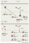
Concomitant activation of proinflammatory transcription factors and the glucocorticoid receptor reinforces inflammation. The ability of the glucocorticoid receptor (GR) to activate gene transcription relies on its ability to interact with glucocorticoid response elements (GREs). (Top) Chromatin, in its inactive state, prevents GR from accessing a GRE and alters the transcriptional response. (Bottom) Proinflammatory transcription factors (TF) promote an open chromatin state in the absence (AP-1) or presence (NK-κB or STAT3) of an inflammatory stimulus, allowing GR to now access chromatin and elicit a response. Some of the glucocorticoid-responsive changes occur as a result of tethering (left) or as a result of a composite regulation (right) where GR is able both to bind to a GRE and tether to the transcription factor. The profile of these complex interactions will shape the immune response and can be either stimulatory or repressive.
Figure 3
Figure 3
Glucocorticoid receptor (GR)-mediated repression of the proinflammatory transcription factors AP-1 and NF-κB. (a) Proinflammatory stimuli trigger a signaling cascade that results in the activation of the transcription factor, AP-1, a heterodimer composed of c-Jun and c-Fos. This drives the transcription of several proinflammatory molecules. Activation of the GR results in its translocation to the nucleus where it can now repress AP-1 activity via one of three mechanisms: (i) at some promoters, GR physically interacts with c-Jun in a process known as tethering, which represses the activity of AP-1 and represses the transcription of proinflammatory genes; (ii) at some promoters, GR is able to simultaneously bind to a GRE and tether to c-Jun to repress the transcriptional activity of AP-1; and (iii) GR induces the expression of MKP-1, a phosphatase, that is able to dephosphorylate and inactivate the kinase JNK. (b) Proinflammatory stimuli trigger a signaling cascade that results in the activation of the transcription factor, NF-κB, a heterodimer composed of the p50 and p65 subunits. This drives the transcription of several proinflammatory genes. Although the exact mechanism is not known, there are several theories as to how GR can inhibit NF-κB activity: (i) similarly to AP-1, GR can physically interact with and repress the activity of NF-κB; (ii) GR is able to block the formation of an NF-κB/IRF3 heterodimer, possibly through the recruitment of GRIP; (iii) GR is able to block the recruitment of the C-terminal tail kinase, pTEFb, thus preventing RNA polymerase II (Pol II) phosphorylation and activation; (iv) GR is able to repress NF-κB activity by recruiting a histone deacetylase (HDAC); (v) GR is able to block the ability of NF-κB to interact with p300 and CPB; and (vi) p53 is able to interact with GR, altering its transcriptional activity, and thus preventing NF-κB activity.
Figure 4
Figure 4
Glucocorticoids promote the resolution of inflammation and restore homeostasis. Glucocorticoids affect nearly every cell type by virtue of nearly ubiquitous expression of the glucocorticoid receptor (GR). During the course of inflammation, glucocorticoids are able to promote resolution by repressing the expression of adhesion molecules, preventing rolling adhesion and extravasation of neutrophils. Glucocorticoids also induce the expression and secretion of Annexin-1, which is able to induce apoptosis of neutrophils at the site of inflammation. Prolonged glucocorticoid exposure induces tissue resident macrophages (MΦ) to undergo a phenotypic change to become M2-like or anti-inflammatory. These macrophages no longer produce proinflammatory cytokines. Instead they produce interleukin-10 (IL-10), have enhanced phagocytic activity to remove apoptotic cells, and promote tissue healing. Glucocorticoids also act on naïve and differentiated T cells that have been recruited to the inflammatory site by blocking T helper 1 (Th1)- and Th2-derived cytokine production as well as inducing apoptosis.
Figure 5
Figure 5
The glucocorticoid receptor (GR) acts a cellular rheostat to ensure the proper response is elicited by the immune system. Glucocorticoids, acting through GR, affect all phases of the immune response. By enhancing the expression of TLR2, P2Y2R, and NLRP3, glucocorticoids ready the innate immune system to respond to microbial products and tissue injury. Additionally, glucocorticoids increase the levels of circulating bone marrow-derived neutrophils. Glucocorticoids reinforce the immune system by cooperating with the proinflammatory transcription factors AP-1, NF-κB, and STAT3. This is accomplished by repressing their activity at specific promoters, inhibiting the production of proinflammatory cytokines. However, GR can also act synergistically with these transcription factors, enhancing the expression and activation of some proinflammatory responses. Glucocorticoids are able to promote the resolution of inflammation, and restore homeostasis, by stimulating the secretion of proresolving molecules (Annexin-1), shifting T cell signaling towards a Th2 response, inducing neutrophil and T cell apoptosis, promoting a wound-healing and antiinflammatory phenotype in macrophages (MΦ), and promoting the removal of apoptotic cells. The ability of GR to accomplish these pleiotropic actions will depend on several factors including post-translational modifications, extracellular environment, ligand availability and duration of signaling, cell type-specific cofactors and binding partners, and chromatin accessibility.

Similar articles

Cited by

References

    1. Newton K, Dixit VM. Signaling in innate immunity and inflammation. Cold Spring Harb Perspect Biol. 2012;4:a006049. - PMC - PubMed
    1. Nathan C, Ding A. Nonresolving inflammation. Cell. 2010;140:871–882. - PubMed
    1. Clark AR, Belvisi MG. Maps and legends: the quest for dissociated ligands of the glucocorticoid receptor. Pharmacol Ther. 2012;134:54–67. - PubMed
    1. Oakley RH, Cidlowski JA. Cellular processing of the glucocorticoid receptor gene and protein: new mechanisms for generating tissue-specific actions of glucocorticoids. J Biol Chem. 2011;286:3177–3184. - PMC - PubMed
    1. Cole TJ, et al. Targeted disruption of the glucocorticoid receptor gene blocks adrenergic chromaffin cell development and severely retards lung maturation. Genes Dev. 1995;9:1608–1621. - PubMed

MeSH terms