Review
. 2013 Mar;19(3):144-56.
doi: 10.1016/j.molmed.2012.12.001. Epub 2013 Jan 9.

Wnt your brain be inflamed? Yes, it Wnt!

Affiliations
Review

Wnt your brain be inflamed? Yes, it Wnt!

Bianca Marchetti et al. Trends Mol Med. 2013 Mar.

Abstract

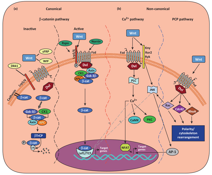
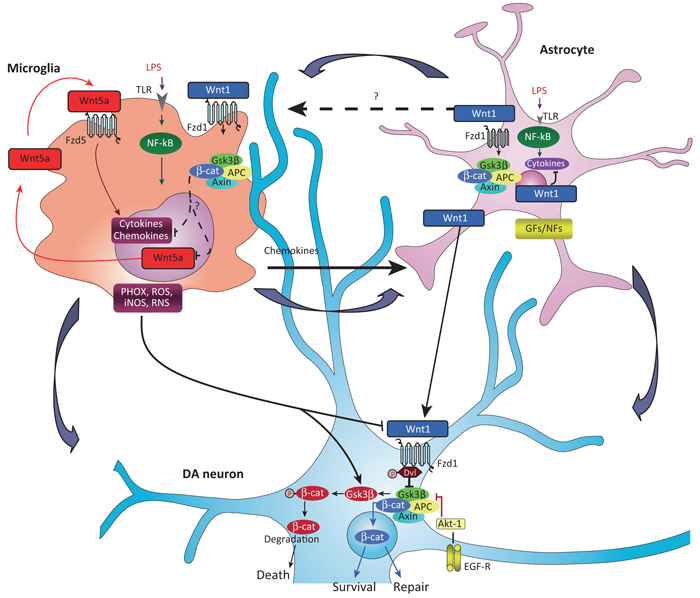
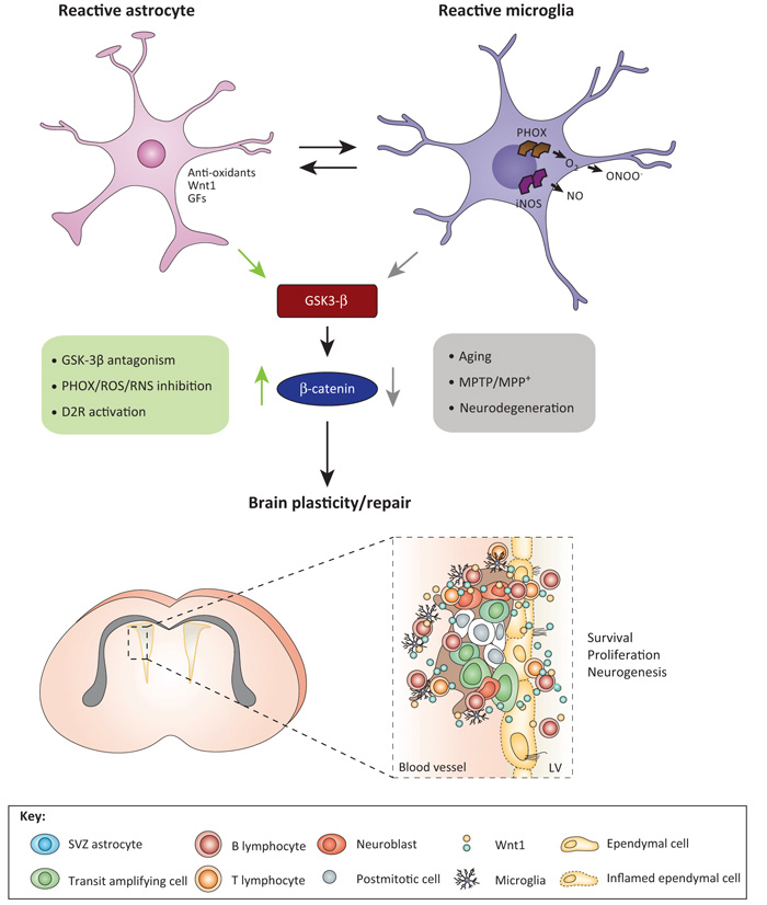
The roles of Wnts in neural development, synaptogenesis, and cancer are generally well characterized. Nonetheless, evidence exists that interactions between the immune and nervous systems control major brain regenerative processes ranging from physiological or pathological (reparative) regeneration to neurogenesis and synaptic plasticity. Recent studies describe deregulated Wnt-Fzd signaling in degenerative and inflammatory central nervous system (CNS) disorders, and the expression of Wnt signaling components in the immune system, and in immune-like cells of the mammalian CNS. This would suggest a likely involvement of Wnts in inflammation-driven brain damage and inflammation-directed brain repair. Here, we review how Wnts modulate neuroimmune interactions and offer a perspective on the most challenging therapeutic opportunities for those CNS diseases where injury-reactive Wnt-flavored inflammation precedes secondary neurodegeneration.


Figures

Figure 1
Figure 1
Canonical or Wnt/β-catenin–TCF/LEF signaling. Three Wnt-dependent pathways have been proposed: canonical Wnt/β-catenin pathway and non-canonical Wnt/PCP and Wnt/Ca2+ pathways. In the canonical Wnt/β-catenin pathway (a), in the absence of Wnt ligands (or in the presence of Wnt inhibitor WIF, sFRPs, or Dkk1), cytosolic β-catenin (β-cat) is targeted to proteolytic degradation through phosphorylation by adenomatous poliposis coli (APC)–Axin–glycogen synthase kinase-3β (GSK-3β)–casein kinase 1 (CK1) destruction complex. Phosphorylated β-cat becomes ubiquitinated through action of β-transducin repeat containing protein–(β-TrCP)-dependent E3 ubiquitin ligase complex and degraded in the proteasome. In the active state, Wnt ligand binding to Fzd receptors and their coreceptors Lrp5/6 leads to the activation of the cytoplasmic protein dishevelled (Dvl), and subsequent recruitment of Axin complex to the Lrp coreceptor, resulting in inhibition of the β-catenin destruction complex. Consequently, hypophosphorylated β-cat accumulate in the cytoplasm and enter the nucleus, where it regulates target gene expression through partnerships with the TCR/LEF1 family of transcription factors, resulting in changes in gene transcription. Two types of proteins, Norrin and R-spondins (Rspo), which are unrelated to Wnt, act as Wnt agonists. In the non-canonical Wnt–Ca2+ signaling pathway (b), the binding of Wnt promotes Fzd-mediated activation of pertussis Toxin-sensitive heterotrimeric guanine nucleotide-binding proteins (G proteins). This, in turn, stimulates the release of Ca2+ from intracellular stores, which leads to the activation of Ca2+-dependent effector molecules. Several Ca2+-sensitive targets – protein kinase C (PKC), Ca2+–calmodulin-dependent protein kinase II (CamKII), and the Ca2+–calmodulin-sensitive protein phosphatase calcineurin – have been identified downstream of the Wnt–Ca2+ pathway. Targets of the Wnt–Ca2+ pathway appear to interact with the Wnt–β-catenin pathway at multiple points. Additionally, Fzd receptors in association with Kny, Ror2, or Ryk receptors can activate JNK, promoting target gene expression through AP-1. In the non-canonical Wnt/PCP pathway, the binding of Wnts activates RhoA/B, Cdc42, or Rac1. Dsh activates Rac1 and Rac1 can also activate JNK, resulting in the NFAT pathway. Abbreviations: Dkk1, Dickkopf 1; Fzd, Frizzled; sFRPs, secreted Frizzled-related proteins; Lrp, low-density lipoprotein receptor-related protein; PCP, planar cell polarity; TCF/LEF, T cell factor/lymphoid enhancer factor; WIF, Wnt inhibitory protein; Wnt, Wingless-related MMTV integration site.
Figure 2
Figure 2
Wnt tripartite regulation in the brain. Macrophage/microglia harbor Fzd5 and Fzd1 subtype receptors and can respond to both non-canonical Wnt5a- and canonical Wnt1-type ligands [9,18,40]. Upon inflammatory challenge with LPS or brain injury, TLRs and IFN-γ-mediated activation, macrophage/microglia produce a panel of proinflammatory cytokines and chemokines, of which Wnt5a constitutes one part of a self-perpetrating cycle, via autocrine Wnt5A/CamKII activation and paracrine stimulation of Th-1 cytokines [40]. Upregulation of microglial PHOX-derived ROS, iNOS-derived NO, and GSK-3β, a known regulator of NF-κB-dependent gene transcription, further exacerbate microglia reaction [8,10]. Chemokine-activated astrocytes respond to microglia by inducing the expression and release of Wnt1-type ligands [8,10,17]. It is proposed that astrocyte-derived Wnt1-type ligands may restrain inflammation, via stimulation of microglia Fzd1 receptors (in analogy to what is observed in macrophages [40]), possibly resulting in the attenuation of cytokines and Wnt5a overexpression (broken arrows). At the neuronal level, MPTP-induced microglial PHOX and RNS upregulate GSK-3β in DA neurons [8,10,17] leading to β-catenin phosphorylation and proteosomal degradation, which may increase DA neuron vulnerability and further incite cell death [8,17]. However, depending on the severity of brain insult, the degree of inflammation and specific vulnerability factors (age, gender, concomitant presence of stressors and gene mutations [87]), astrocyte–neuron crosstalk, via Wnt1 may serve a protective role, via stabilization of β-catenin in the cytoplasm, its nuclear translocation, followed by transcription of prosurvival genes, that may lead to neuroprotection and/or neurorepair [8,17]. Chemokine-activated astrocytes can also promote neurogenesis from adult VM progenitors, in vitro, via Wnt/β-catenin signaling activation [8]. In stark contrast, aging-induced loss of astrocyte-derived Wnt1 response [8,15], or Wnt/β-catenin signaling antagonism with Dkk1 [17], may result in DA neuron failure to repair. Potential crosstalk between glial GFs/NFs and Wnt/β-catenin signaling via Akt/GSK-3β/β-catenin cascades are also illustrated (see [17]). Abbreviations: CamKII, Ca2+–calmodulin-dependent protein kinase II; DA, dopamine; Dkk1, Dickkopf 1; EGF, epidermal growth factor; Fzd, Frizzled; GFs/NFs, glial-derived growth/neurotrophic factors; IFN-γ, interferon-γ; LPS, lipopolysaccharide; MPTP, 1-methyl-4-phenyl-1,2,3,6-tetrahydropyridine; NF-κB, nuclear factor (NF)-κB; PHOX, phagocyte oxidase; RNS, reactive nitrite species; ROS, reactive oxygen species; Th-1, T helper 1 cells; TLR, Toll-like receptor; VM, ventral midbrain; Wnt, Wingless-related MMTV integration site.
Figure 3
Figure 3
Crosstalk between Wnt and local inflammatory signaling pathways for the control of SVZ niche homeostasis. During the early degeneration phase of MPTP toxicity, hyperactivated microglia contribute to the impairment of SVZ neurogenesis at different levels. By increasing oxidative and nitrosative stress and in synergy with MPTP/MPP+ direct toxicity, microglial-derived mediators (PHOX-derived ROS and iNOS-derived NO and peroxynitrite) may act as a molecular switch for cell signaling pathways critically involved in the physiological control of NPC homeostasis, with harmful consequences for NPC physiology, at least in part through GSK-3β activation, followed by phosphorylation and consequent degradation of β-catenin [10,15]. By contrast, pharmacological mitigation of inflammation and oxidative stress with Apo, L-Nil, or HCT1026 upregulate β-catenin and successfully rescue NPC proliferation and neuroblast formation, a process associated with striatal DAergic neuroprotection, with further positive modulation of SVZ proliferation via D2-R-activated mechanisms [10]. The mutual role of astrocyte–microglial interactions in the plasticity of SVZ response to MPTP is exemplified by the astrocyte’s ability to overcome microglial inhibitory effects, through, besides others, Wnt1-mediated activation Wnt/β-catenin signaling [10]. Different upstream and downstream signaling cascades may converge in finely tuning β-catenin transcriptional activity. For example, NO can inhibit EGF-R and the phosphoinositide 3-kinases (PI3K)/AKT survival pathway, and GSK-3β is a downstream target of Akt; RNS-induced nitration regulates the p85 subunit of PI3 kinase; and a variety of molecules including growth factors and neurotransmitter, including DA via D2 receptor can signal through (PI3K)/AKT/GSK-3β and Wnt signaling activation, thus, crosstalk among Wnt/β-catenin and prominent intracellular pathways may be envisioned to fine tune the SVZ neurogenic potential/plasticity observed herein [10]. Crosstalk between SVZ astrocytes (blue), transit-amplifying cells (red), neuroblasts (orange), ependymal (yellow) cells, microglia (violet), and lymphocytes via Wnt in SVZ niche are schematically illustrated. Abbreviations: Apo, apocynin, a ROS antagonist; DA, dopamine; D2-R, dopaminergic receptor subtype 2; HCT1026, [2-fluoro-α-methyl(1,1′-biphenyl)-4-acetic-4-(nitrooxy)butyl ester], a mixed cyclooxygenase (COX1/COX2) inhibitor NO-donating non-steroidal anti-inflammatory drug (NSAID) endowed with additional anti-inflammatory activity and reduced side effects; L-Nil, L-N6-(1-iminoethyl)-lysine, a specific inhibitor of inducible nitric oxide synthase (iNOS); MPP+, 1-methyl-4-phenylpyridinium; MPTP, 1-methyl-4-phenyl-1,2,3,6-tetrahydropyridine; NPC, neural stem/progenitor cell; PHOX, phagocyte oxidase; RNS, reactive nitrogen species; ROS, reactive oxygen species; SVZ, subventricular zone. Adapted from [111].

Similar articles

Cited by

References

    1. Salinas PC. Wnt signaling in the vertebrate central nervous system: from axon guidance to synaptic function. Cold Spring Harb. Perspect. Biol. 2012;4:a008003. - PMC - PubMed
    1. Willert K, Nusse R. Wnt proteins. Cold Spring Harb. Perspect. Biol. 2012;4:a007864. - PMC - PubMed
    1. Harrison-Uy SJ, Pleasure SJ. Wnt signaling and forebrain development. Cold Spring Harb. Perspect. Biol. 2012;4:a008094. - PMC - PubMed
    1. Toledo EM, et al. Wnt signaling in neuroprotection and stem cell differentiation. Prog. Neurobiol. 2008;86:281–296. - PubMed
    1. Mastroiacovo F, et al. Induction of the Wnt antagonist, Dickkopf-1, contributes to the development of neuronal death in models of brain focal ischemia. J. Cereb. Blood Flow Metab. 2009;29:264–276. - PubMed

Publication types

LinkOut - more resources