. 2013 Jun:54:206-15.
doi: 10.1016/j.nbd.2012.12.013. Epub 2013 Jan 8.

Astrocyte GRK2 as a novel regulator of glutamate transport and brain damage

Affiliations

Astrocyte GRK2 as a novel regulator of glutamate transport and brain damage

Cora H Nijboer et al. Neurobiol Dis. 2013 Jun.

Abstract

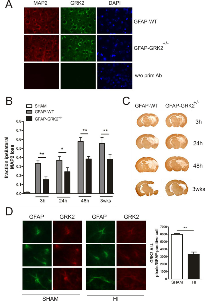
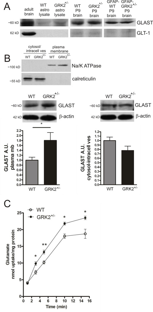
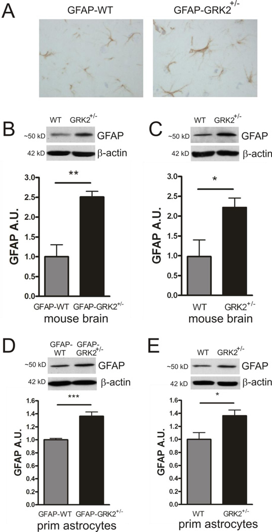
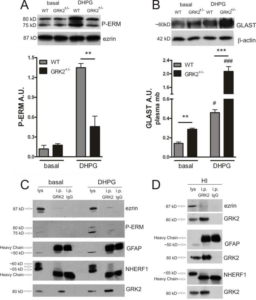
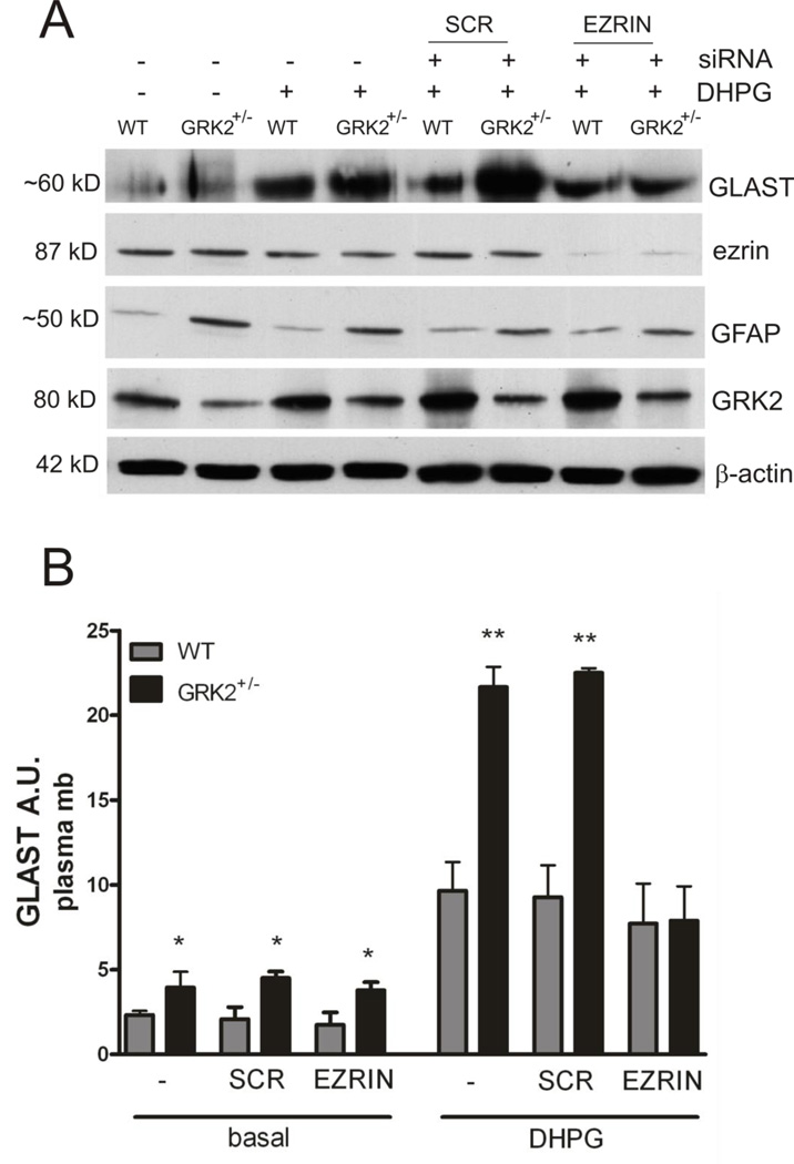
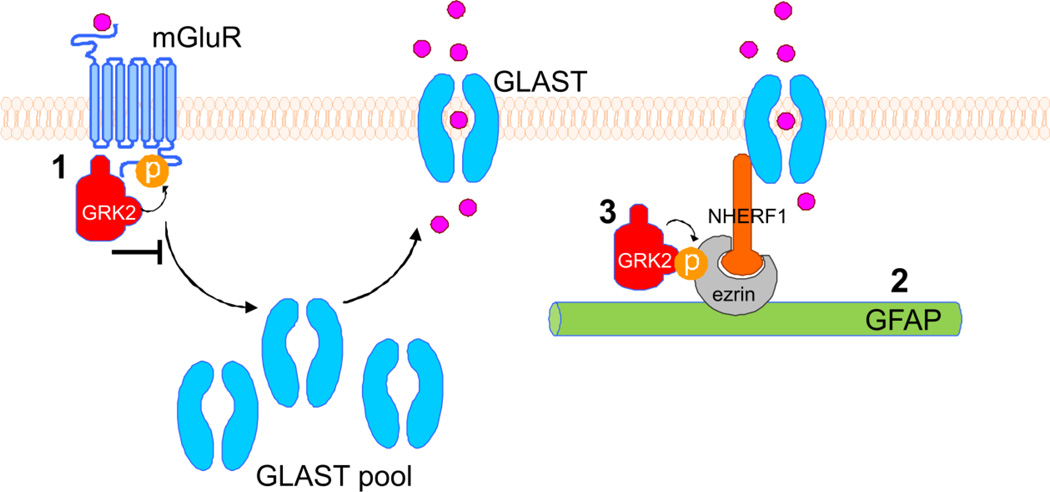
G protein-coupled receptor (GPCR) kinase 2 (GRK2) regulates cellular signaling via desensitization of GPCRs and by direct interaction with intracellular signaling molecules. We recently described that ischemic brain injury decreases cerebral GRK2 levels. Here we studied the effect of astrocyte GRK2-deficiency on neonatal brain damage in vivo. As astrocytes protect neurons by taking up glutamate via plasma-membrane transporters, we also studied the effect of GRK2 on the localization of the GLutamate ASpartate Transporter (GLAST). Brain damage induced by hypoxia-ischemia was significantly reduced in GFAP-GRK2(+/-) mice, which have a 60% reduction in astrocyte GRK2 compared to GFAP-WT littermates. In addition, GRK2-deficient astrocytes have higher plasma-membrane levels of GLAST and an increased capacity to take up glutamate in vitro. In search for the mechanism by which GRK2 regulates GLAST expression, we observed increased GFAP levels in GRK2-deficient astrocytes. GFAP and the cytoskeletal protein ezrin are known regulators of GLAST localization. In line with this evidence, GRK2-deficiency reduced phosphorylation of the GRK2 substrate ezrin and enforced plasma-membrane GLAST association after stimulation with the group I mGluR-agonist DHPG. When ezrin was silenced, the enhanced plasma-membrane GLAST association in DHPG-exposed GRK2-deficient astrocytes was prevented. In conclusion, we identified a novel role of astrocyte GRK2 in regulating plasma-membrane GLAST localization via an ezrin-dependent route. We demonstrate that the 60% reduction in astrocyte GRK2 protein level that is observed in GFAP-GRK2(+/-) mice is sufficient to significantly reduce neonatal ischemic brain damage. These findings underline the critical role of GRK2 regulation in astrocytes for dampening the extent of brain damage after ischemia.


Figures

Figure 1
Figure 1. Functional consequences of astrocyte GRK2-deficiency for brain damage in vivo
A: Representative photographs of immunofluorescent staining of MAP2 (red), GRK2 (green) and DAPI (blue) in the cortex of GFAP-WT and GFAP-GRK2+/− P9 mice, illustrating neuronal GRK2 levels. As a control the primary antibodies were ommitted (w/o prim Ab). B: Quantification of HI-induced MAP2 loss in the ipsilateral hemisphere at hippocampal level at 3 h, 24 h, 48 h and 3 wks post- HI in GFAP-WT and GFAP-GRK2+/− animals and in sham-operated controls. MAP2 loss was calculated as 1- {MAP2-positive staining in ipsilateral/contralateral hemisphere}. Sham-operated controls of both genotypes did not show significant MAP2 loss and are represented as one group. n=10-12 per genotype per time point. C: Representative photographs of immunohistochemical staining of MAP2 as an indicator of neuronal integrity in GFAP-WT and GFAP-GRK2+/− mice at the different time points indicated. D: Left: Representative photographs of immunofluorescent staining of GFAP (green) and GRK2 (red) in the white matter tract of sham-operated (SHAM) or HI-treated (HI) P9 mice at 3 h post-HI. Right: quantification of GRK2 level in GFAP-positive cells. *p<0.05; **p<0.01 GFAP-WT vs GFAP-GRK2+/−; **p<0.01 SHAM vs HI.
Figure 2
Figure 2. Functional consequences of low astrocyte GRK2 for plasma-membrane GLAST expression and glutamate uptake
A: Western Blot examples showing expression of GLAST and not GLT-1 in total lysates of primary cultures of WT and GRK2-deficient astrocytes. Total protein samples of adult WT and P9 WT and GRK2+/− or P9 GFAP-WT and GFAP-GRK2+/− mouse brains are loaded as controls. B: Upper part shows quality of subcellular fractionation of astrocyte lysates into plasma-membrane fraction and fraction containing cytosol and intracellular vesicular membranes (cytosol/intracell ves). Note exclusive presence of Na+/K+ ATPase in plasma-membrane fraction and exclusive presence of calreticulin, a marker for ER vesicles, in the cytosol/intracellular vesicle fraction. Lower part shows GLAST expression in plasma-membrane (plasma mb) fraction (left) and cytosol/intracellular vesicle fraction (right) of WT and GRK2-deficient primary cultures of astrocytes analyzed by Western Blotting. Insets show representative Western Blot examples; β-actin is used as a loading control for both fractions. Ponceau S staining gave similar equal loading results as observed after probing with β-actin antibody. n=8 animals per genotype. C: Glutamate uptake capacity of WT and GRK2-deficient astrocytes was tested by measuring 3H-labeled glutamate (100 µM) uptake over 15 minutes. Data are presented as nmol glutamate uptake per mg protein. n=9 per genotype per time point. A.U.: arbitrary units. *p<0.05; **p<0.01 WT vs GRK2+/−.
Figure 3
Figure 3. GRK2-deficiency is associated with increased GFAP in astrocytes
A: Representative photographs of immunohistochemical staining of GFAP illustrating increased GFAP expression in parietal cortices of sham-control GFAP-GRK2+/− mice compared to GFAP-WT littermates. B–C: GFAP expression in total brain homogenates of GFAP-WT and GFAP-GRK2+/− (B) or WT and GRK2+/− (C) mice analyzed by Western Blotting. Insets show representative Western Blot examples; β-actin is used as a loading control. n=5-6 animals per genotype. D–E: GFAP expression in total lysates of primary cultures of GFAP-WT and GFAP-GRK2+/− (D)or WT and GRK2+/− (E) astrocytes analyzed by Western Blotting. Insets show representative Western Blot examples; β-actin is used as loading control. n=6-8 animals per genotype. A.U.: arbitrary units. *p<0.05; ***p<0.001 WT vs GRK2+/− or GFAP-WT vs GFAP-GRK2+/−
Figure 4
Figure 4. GRK2 deficiency decreases DHPG-induced P-ERM and increases DHPG-induced plasma-membrane GLAST association
A: Phosphorylated ezrin (P-ERM) in plasma-membrane fractions of WT and GRK2+/− astrocytes after 10 min of stimulation with culture medium (basal) or DHPG (50 µM) analyzed by Western Blotting. Insets show representative Western Blot examples; total ezrin is used as a loading control. **p<0.01 WT vs GRK2+/−. B: GLAST expression in plasma-membrane fractions of WT and GRK2+/− astrocytes after 10 min of stimulation with culture medium (basal) or DHPG (50 µM) analyzed by Western Blotting. Insets show representative Western Blot examples; β-actin is used as a loading control. # p<0.05, ### p<0.001: basal vs DHPG per genotype. **p<0.01; ***p<0.001: WT vs GRK2+/−. A/B : Data are from 3 independent experiments (n=3) performed in 8-fold. C–D: GRK2 protein was immunoprecipitated (i.p. GRK2) in samples from unstimulated (basal) and DHPG-stimulated cultures of primary WT astrocytes (C) or in P9 mouse brain homogenates pooled from 5 WT animals at 0.5 h post-HI (D). The same amount of mouse IgG (IgG2a kappa) was used as a control (i.p. IgG). The immunoprecipitated products were analyzed by Western Blotting. Blots show co-immunoprecipitation of ezrin, phosphorylated ezrin (P-ERM) (after DHPG in cultured astrocytes), GFAP and NHERF1 with GRK2. Arrows indicate migration of the bands of interest and the heavy chain of the IgG around 50 kD. Total astrocyte lysate (lys; C) or total brain homogenate (lys; D) (10% of input) was loaded for comparison. GRK2 blots show increased levels of GRK2 after immunoprecipitation, indicating successful immunoprecipitation.
Figure 5
Figure 5. Increased DHPG-induced GLAST association with the plasma-membrane in GRK2-deficient cells is dependent on ezrin
A: Ezrin protein expression was silenced in WT and GRK2+/− astrocytes using siRNA. 48 h later, astrocytes were stimulated with culture medium (basal) or DHPG (50 µM for 10 min) and protein lysates were analyzed by Western Blotting. β-actin is used as a loading control. Ezrin protein level was reduced >90% after transfection with siRNA. Transfection with scrambled (SCR) siRNA did not have any effect on ezrin protein levels. B: Quantification of GLAST expression in plasma-membrane fractions of WT and GRK2+/− astrocytes after siRNA transfection (ezrin or scrambled) and stimulation with culture medium (basal) or DHPG (see A). Data are from 2-4 independent experiments performed in 4-fold. *p<0.05; **p<0.01
Figure 6
Figure 6. Diagram showing possible regulatory mechanisms of GRK2 on GLAST plasma-membrane localization
Left part of diagram: upon binding of glutamate or a mGluR agonist, mGluRs are activated and start signaling. As a consequence, GLAST is redistributed from cytoplasmic pools to the plasma-membrane, where the GLAST transporters take up glutamate. Our results show that GRK2-deficient astrocytes express higher levels of plasma-membrane GLAST under basal and stimulated (mGluR-agonist DHPG) conditions. Several mechanisms for a regulatory role of GRK2 can be proposed: 1: GRK2 phosphorylates group I mGluRs, leading to termination of signaling downstream of these receptor and receptor internalization (inhibitory arrow). GRK2-deficiency leads to enhanced signaling via the mGluR and might directly regulate GLAST distribution. 2: A crucial role for GFAP intermediate filaments has been shown for retainment of GLAST in the plasma-membrane (Sullivan et al., 2007). GRK2-deficient astrocytes show enhanced GFAP expression, which might lead to enhanced linking of GLAST in the plasma-membrane. 3: GFAP regulates GLAST plasma-membrane localization via linker proteins ezrin and NHERF1. Ezrin is a substrate of GRK2. Stimulation of the mGluR induces phosphorylation of ezrin. GRK2-deficient astrocytes show reduced phospho-ezrin levels. Silencing of ezrin completely abolished the increase GLAST plasma-membrane expression in GRK2-deficient astrocytes. Our data show that DHPG-induced regulation of GLAST transport to the plasma-membrane becomes dependent on ezrin only when GRK2 is low.

Similar articles

Cited by

References

    1. Beaulé C, Swanstrom A, Leone MJ, Herzog ED. Circadian modulation of gene expression, but not glutamate uptake, in mouse and rat cortical astrocytes. PLoS One. 2009;4:e7476. - PMC - PubMed
    1. Cant SH, Pitcher JA. G protein-coupled receptor kinase 2-mediated phosphorylation of ezrin is required for G protein-coupled receptor-dependent reorganization of the actin cytoskeleton. Mol Biol Cell. 2005;16:3088–3099. - PMC - PubMed
    1. Dale LB, Bhattacharya M, Anborgh PH, Murdoch B, Bhatia M, Nakanishi S, Ferguson SS. G protein-coupled receptor kinasemediated desensitization of metabotropic glutamate receptor 1A protects against cell death. J Biol Chem. 2000;275:38213–38220. - PubMed
    1. Danbolt NC. Glutamate uptake. Prog Neurobiol. 2001;65:1–105. - PubMed
    1. Duan S, Anderson CM, Stein BA, Swanson RA. Glutamate induces rapid upregulation of astrocyte glutamate transport and cellsurface expression of GLAST. J Neurosci. 1999;19:10193–10200. - PMC - PubMed

Publication types