

Noninvasive detection of fetal subchromosome abnormalities via deep sequencing of maternal plasma
- PMID: 23313373
- PMCID: PMC3567270
- DOI: 10.1016/j.ajhg.2012.12.006
Noninvasive detection of fetal subchromosome abnormalities via deep sequencing of maternal plasma
Abstract
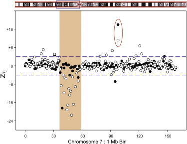
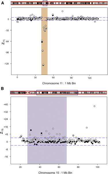
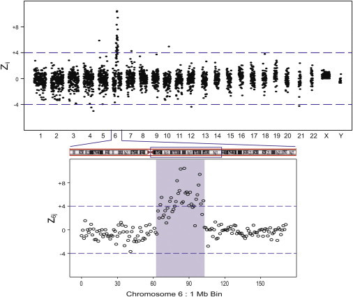
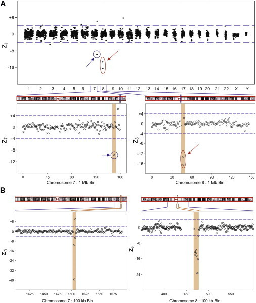
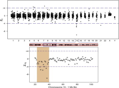
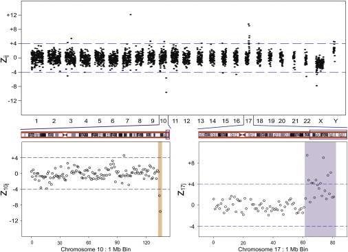
The purpose of this study was to determine the deep sequencing and analytic conditions needed to detect fetal subchromosome abnormalities across the genome from a maternal blood sample. Cell-free (cf) DNA was isolated from the plasma of 11 pregnant women carrying fetuses with subchromosomal duplications and deletions, translocations, mosaicism, and trisomy 20 diagnosed by metaphase karyotype. Massively parallel sequencing (MPS) was performed with 25-mer tags at approximately 10(9) tags per sample and mapped to reference human genome assembly hg19. Tags were counted and normalized to fixed genome bin sizes of 1 Mb or 100 kb to detect statistically distinct copy-number changes compared to the reference. All seven cases of microdeletions, duplications, translocations, and the trisomy 20 were detected blindly by MPS, including a microdeletion as small as 300 kb. In two of these cases in which the metaphase karyotype showed additional material of unknown origin, MPS identified both the translocation breakpoint and the chromosomal origin of the additional material. In the four mosaic cases, the subchromosomal abnormality was not demonstrated by MPS. This work shows that in nonmosaic cases, it is possible to obtain a fetal molecular karyotype by MPS of maternal plasma cfDNA that is equivalent to a chromosome microarray and in some cases is better than a metaphase karyotype. This approach combines the advantage of enhanced fetal genomic resolution with the improved safety of a noninvasive maternal blood test.
Copyright © 2013 The American Society of Human Genetics. Published by Elsevier Inc. All rights reserved.
Figures
References
-
- Miller D.T., Adam M.P., Aradhya S., Biesecker L.G., Brothman A.R., Carter N.P., Church D.M., Crolla J.A., Eichler E.E., Epstein C.J. Consensus statement: chromosomal microarray is a first-tier clinical diagnostic test for individuals with developmental disabilities or congenital anomalies. Am. J. Hum. Genet. 2010;86:749–764. - PMC - PubMed
-
- Vetro A., Bouman K., Hastings R., McMullan D.J., Vermeesch J.R., Miller K., Sikkema-Raddatz B., Ledbetter D.H., Zuffardi O., van Ravenswaaij-Arts C.M. The introduction of arrays in prenatal diagnosis: a special challenge. Hum. Mutat. 2012;33:923–929. - PubMed
-
- Novelli A., Grati F.R., Ballarati L., Bernardini L., Bizzoco D., Camurri L., Casalone R., Cardarelli L., Cavalli P., Ciccone R. Microarray application in prenatal diagnosis: a position statement from the cytogenetics working group of the Italian Society of Human Genetics (SIGU), November 2011. Ultrasound Obstet. Gynecol. 2012;39:384–388. - PubMed
Publication types
MeSH terms
Substances
LinkOut - more resources
Full Text Sources
Other Literature Sources
Miscellaneous
