. 2013 Feb 7;92(2):259-64.
doi: 10.1016/j.ajhg.2012.12.002. Epub 2013 Jan 11.

LRIG2 mutations cause urofacial syndrome

Affiliations

LRIG2 mutations cause urofacial syndrome

Helen M Stuart et al. Am J Hum Genet. .

Abstract

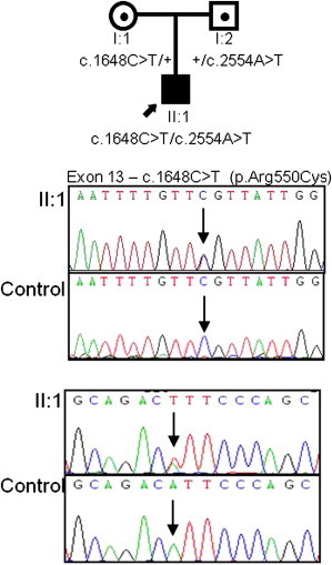
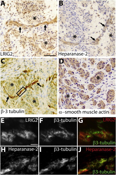
Urofacial syndrome (UFS) (or Ochoa syndrome) is an autosomal-recessive disease characterized by congenital urinary bladder dysfunction, associated with a significant risk of kidney failure, and an abnormal facial expression upon smiling, laughing, and crying. We report that a subset of UFS-affected individuals have biallelic mutations in LRIG2, encoding leucine-rich repeats and immunoglobulin-like domains 2, a protein implicated in neural cell signaling and tumorigenesis. Importantly, we have demonstrated that rare variants in LRIG2 might be relevant to nonsyndromic bladder disease. We have previously shown that UFS is also caused by mutations in HPSE2, encoding heparanase-2. LRIG2 and heparanase-2 were immunodetected in nerve fascicles growing between muscle bundles within the human fetal bladder, directly implicating both molecules in neural development in the lower urinary tract.


Figures

Figure 1
Figure 1
Identification of Mutations in LRIG2 in Three Families Affected by UFS Pedigrees and LRIG2 mutation analysis in families 1 (A), 2 (B), and 3 (C). (A) In family 1, a homozygous frameshift (c.1230delA [p.Glu410Aspfs6]) in exon 10 was identified. (B) In family 2, affected child II-I is shown at the age of 6 months. Her voiding cystourethrogram shows a trabeculated bladder and severe left-sided VUR (Bi). A compound-heterozygous frameshift (c.2088delC [p.Ser697Hisfs11]) (Bii) and a compound-heterozygous AluYa5-like insertion (c.1980_1981ins371, GenBank JX891452) (Biii) were identified. Gel electrophoresis demonstrated a larger band present in I:2, II:1, and II:2, and sequencing of cDNA confirmed the heterozygous skipping of exon 14 in the same individuals. (C) In family 3, a homozygous nonsense mutation in exon 15 (c.2125C>T [p.Arg709]) was identified. All individuals genotyped for LRIG2 variants are indicated by an asterisk. The mutations segregated in the three families consistently with the phenotype.
Figure 2
Figure 2
Identification of Mutations in LRIG2 in an Individual with Nonsyndromic Dysfunctional Voiding Pedigree and LRIG2 mutation analysis in this family revealed compound heterozygous missense mutations, c.1648C>T [p.Arg550Cys] (exon 13) and c.2554A>T [p.Ile852Phe] (exon 16), in affected individual II:1.
Figure 3
Figure 3
Immunohistochemistry of a Normal Urinary Bladder at 12 Weeks of Gestation (A–D) Bright-field images counterstained with hematoxylin (blue nuclei). (A) Immunodetection of LRIG2 (brown). Note the prominent signal in a linear structure (arrows) between muscle bundles (asterisks), which themselves show weaker LRIG2 immunoreactivity. The scale bar represents 50 μm. (B) Heparanase-2 immunodetected in a nerve-like structure between muscle bundles. (C) The linear structure contains components with β3-tubulin immunoreactivity, an axonal protein, thus confirming that it is a nerve. (D) Detrusor muscle bundles express α-smooth muscle actin. (E–J) High-power dark-field images of an area of nerve similar to that in the outlined box in (C). LRIG2 and β3-tubulin immunoreactivity (white) are depicted in (E) and (F), respectively. The color merged image (G) shows that the signal for LRIG2 (red) is generally discrete from the signal (green) for β3-tubulin; given that the latter is an axonal marker, LRIG2 might be mainly localized in neural support cells, the exact nature of which remains to be established. Heparanse-2 and β3-tubulin immunoreactivity (white) are depicted in (H) and (I), respectively. The merged image (J) shows that the signal for heparanase-2 partially overlaps with that of β3-tubulin to generate a yellow color. Histology, incorporating 5 μm paraffin sections of paraformaldehyde-fixed tissues, was undertaken with human fetal tissue collected with ethical approval under the Codes of Practice of the UK Human Tissue Authority. Endogenous peroxidise was quenched by incubation with hydrogen peroxide, and antigen retrieval was undertaken by heating at 95°C for 5 minutes in sodium citrate (pH 6). The primary antibodies used were goat anti-α-smooth muscle actin (Sigma-Aldrich SAB2500963), chicken anti-β3-tubulin (Millipore AB9354), rabbit anti-LRIG2 (Abgent AP13821b), and rabbit anti-heparanase-2 (Generon). The latter was generated against a unique epitope (NH2-QLDPSIIHDGWLDC-CONH2). After application of appropriate secondary antibodies, a streptavidin-horseradish-peroxidase-DAB system was used. For coimmunostaining immunofluorescence studies, the quenching step was omitted and species-specific secondary antibodies, each conjugated to a different fluorophores with nonoverlapping emission spectra, were used for detecting the proteins under study. The scale bar in (E) represents 10 μm.

References

    1. Kerecuk L., Schreuder M.F., Woolf A.S. Renal tract malformations: Perspectives for nephrologists. Nat. Clin. Pract. Nephrol. 2008;4:312–325. - PubMed
    1. Adalat S., Bockenhauer D., Ledermann S.E., Hennekam R.C., Woolf A.S. Renal malformations associated with mutations of developmental genes: Messages from the clinic. Pediatr. Nephrol. 2010;25:2247–2255. - PMC - PubMed
    1. Lambert H.J., Stewart A., Gullett A.M., Cordell H.J., Malcolm S., Feather S.A., Goodship J.A., Goodship T.H., Woolf A.S., UK VUR Study Group Primary, nonsyndromic vesicoureteric reflux and nephropathy in sibling pairs: A United Kingdom cohort for a DNA bank. Clin. J. Am. Soc. Nephrol. 2011;6:760–766. - PMC - PubMed
    1. Benarroch E.E. Neural control of the bladder: Recent advances and neurologic implications. Neurology. 2010;75:1839–1846. - PubMed
    1. Weber S., Thiele H., Mir S., Toliat M.R., Sozeri B., Reutter H., Draaken M., Ludwig M., Altmüller J., Frommolt P. Muscarinic acetylcholine receptor M3 mutation causes urinary bladder disease and a prune-belly-like syndrome. Am. J. Hum. Genet. 2011;89:668–674. - PMC - PubMed

Publication types

Substances

Supplementary concepts

Associated data