

Osterix is required for cranial neural crest-derived craniofacial bone formation
- PMID: 23313488
- PMCID: PMC4012829
- DOI: 10.1016/j.bbrc.2012.12.138
Osterix is required for cranial neural crest-derived craniofacial bone formation
Abstract
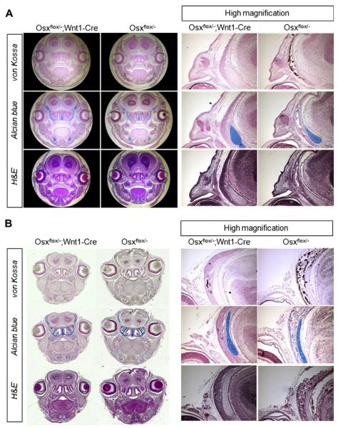
Osx plays essential roles in regulating osteoblast and chondrocyte differentiation, and bone formation during mouse skeletal development. However, many questions remain regarding the requirement for Osx in different cell lineages. In this study, we asked whether Osx is required for craniofacial bone formation derived from cranial neural crest (CNC) cells. The Osx gene was conditionally inactivated in CNC-derived cells using a Wnt1-Cre recombination system. Neural crest-specific inactivation of Osx resulted in the complete absence of intramembranous skeletal elements derived from the CNC, and CNC-derived endochondral skeletal elements were also affected by Osx inactivation. Interestingly, Osx inactivated CNC-derived cells, which were recapitulated by lacZ expression, occupied the same regions of craniofacial skeletal elements as observed for controls. However, cells lost their osteogenic ability to differentiate into functional osteoblasts by Osx inactivation. These results suggest that Osx is important for craniofacial bone formation by CNC-derived cells. This finding provides novel insights of the regulation of craniofacial development by the gene network and transcription factors, and the understanding of human diseases caused by neural crest developmental abnormalities.
Copyright © 2013 Elsevier Inc. All rights reserved.
Figures


References
-
- Huang X, Saint-Jeannet JP. Induction of the neural crest and the opportunities of life on the edge. Dev. Biol. 2004;275:1–11. - PubMed
-
- Chai Y, Jiang X, Ito Y, Bringas P, Jr., Han J, Rowitch DH, Soriano P, McMahon AP, Sucov HM. Fate of the mammalian cranial neural crest during tooth and mandibular morphogenesis. Development. 2000;127:1671–1679. - PubMed
-
- Cerny R, Lwigale P, Ericsson R, Meulemans D, Epperlein HH, Bronner-Fraser M. Developmental origins and evolution of jaws: new interpretation of “maxillary” and “mandibular”. Dev. Biol. 2004;276:225–236. - PubMed
-
- Jiang X, Sachiko I, Maxson RE, Sucov HM, Morriss-Kay GM. Tissue origins and interactions in the mammalian skull vault. Dev. Biol. 2002;241:106–116. - PubMed
Publication types
MeSH terms
Substances
Grants and funding
LinkOut - more resources
Full Text Sources
Other Literature Sources
Medical
Molecular Biology Databases
