

Prolonged diet induced obesity has minimal effects towards brain pathology in mouse model of cerebral amyloid angiopathy: implications for studying obesity-brain interactions in mice
- PMID: 23313575
- PMCID: PMC4566932
- DOI: 10.1016/j.bbadis.2013.01.002
Prolonged diet induced obesity has minimal effects towards brain pathology in mouse model of cerebral amyloid angiopathy: implications for studying obesity-brain interactions in mice
Abstract
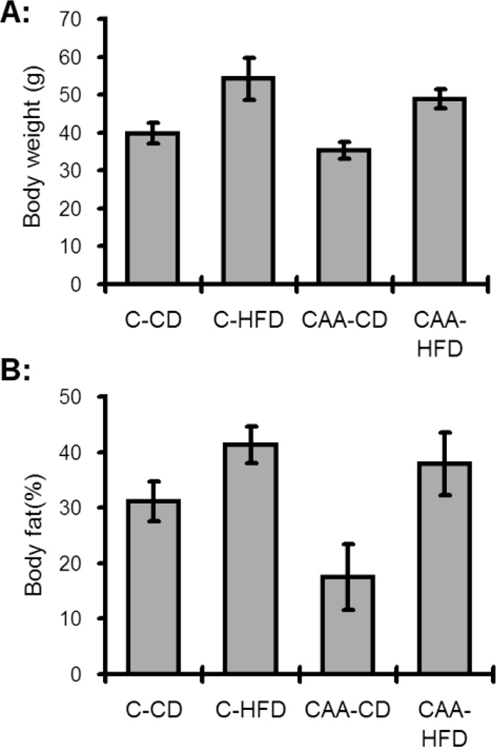
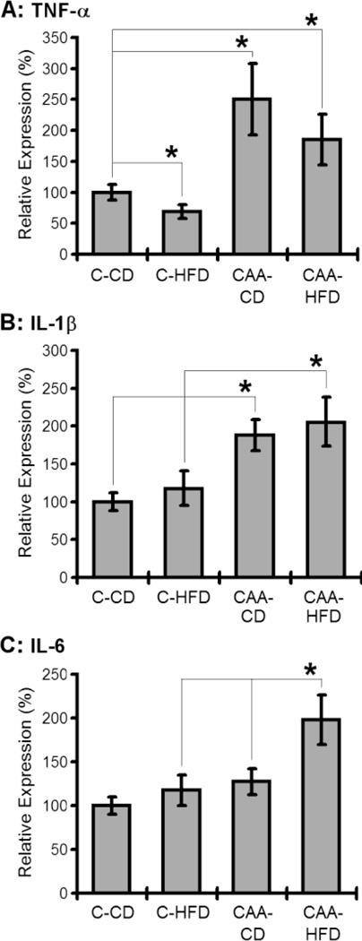
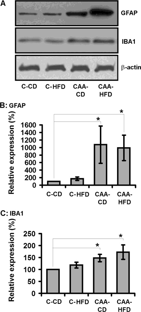
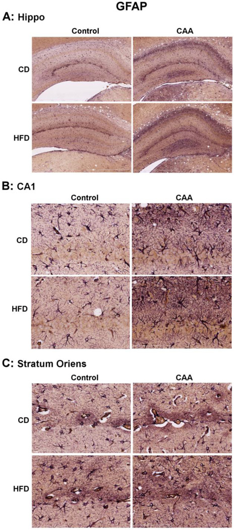
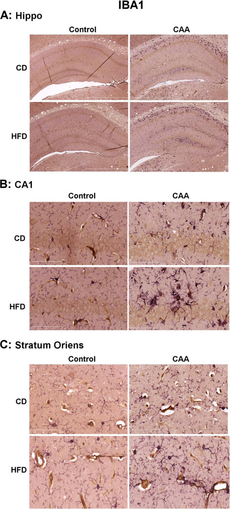
Cerebral amyloid angiopathy (CAA) occurs in nearly every individual with Alzheimer's disease (AD) and Down's syndrome, and is the second largest cause of intracerebral hemorrhage. Mouse models of CAA have demonstrated evidence for increased gliosis contributing to CAA pathology. Nearly two thirds of Americans are overweight or obese, with little known about the effects of obesity on the brain, although increasingly the vasculature appears to be a principle target of obesity effects on the brain. In the current study we describe for the first time whether diet induced obesity (DIO) modulates glial reactivity, amyloid levels, and inflammatory signaling in a mouse model of CAA. In these studies we identify surprisingly that DIO does not significantly increase Aβ levels, astrocyte (GFAP) or microglial (IBA-1) gliosis in the CAA mice. However, within the hippocampal gyri a localized increase in reactive microglia were increased in the CA1 and stratum oriens relative to CAA mice on a control diet. DIO was observed to selectively increase IL-6 in CAA mice, with IL-1β and TNF-α not increased in CAA mice in response to DIO. Taken together, these data show that prolonged DIO has only modest effects towards Aβ in a mouse model of CAA, but appears to elevate some localized microglial reactivity within the hippocampal gyri and selective markers of inflammatory signaling. These data are consistent with the majority of the existing literature in other models of Aβ pathology, which surprisingly show a mixed profile of DIO effects towards pathological processes in mouse models of neurodegenerative disease. The importance for considering the potential impact of ceiling effects in pathology within mouse models of Aβ pathogenesis, and the current experimental limitations for DIO in mice to fully replicate metabolic dysfunction present in human obesity, are discussed. This article is part of a Special Issue entitled: Animal Models of Disease.
Copyright © 2012. Published by Elsevier B.V.
Figures
Similar articles
-
Chronic treatment with five vascular risk factors causes cerebral amyloid angiopathy but no Alzheimer pathology in C57BL6 mice.Brain Behav Immun. 2019 May;78:52-64. doi: 10.1016/j.bbi.2019.01.009. Epub 2019 Jan 19. Brain Behav Immun. 2019. PMID: 30664922
-
ApoA-I deficiency increases cortical amyloid deposition, cerebral amyloid angiopathy, cortical and hippocampal astrogliosis, and amyloid-associated astrocyte reactivity in APP/PS1 mice.Alzheimers Res Ther. 2019 May 13;11(1):44. doi: 10.1186/s13195-019-0497-9. Alzheimers Res Ther. 2019. PMID: 31084613 Free PMC article.
-
A1 reactive astrocytes and a loss of TREM2 are associated with an early stage of pathology in a mouse model of cerebral amyloid angiopathy.J Neuroinflammation. 2020 Jul 25;17(1):223. doi: 10.1186/s12974-020-01900-7. J Neuroinflammation. 2020. PMID: 32711525 Free PMC article.
-
Cerebral amyloid angiopathy and Alzheimer disease - one peptide, two pathways.Nat Rev Neurol. 2020 Jan;16(1):30-42. doi: 10.1038/s41582-019-0281-2. Epub 2019 Dec 11. Nat Rev Neurol. 2020. PMID: 31827267 Free PMC article. Review.
-
Acquired cerebral amyloid angiopathy: An emerging concept.Prog Mol Biol Transl Sci. 2019;168:85-95. doi: 10.1016/bs.pmbts.2019.05.012. Epub 2019 Jun 18. Prog Mol Biol Transl Sci. 2019. PMID: 31699330 Review.
Cited by
-
Obesity and diabetes cause cognitive dysfunction in the absence of accelerated β-amyloid deposition in a novel murine model of mixed or vascular dementia.Acta Neuropathol Commun. 2014 Jun 10;2:64. doi: 10.1186/2051-5960-2-64. Acta Neuropathol Commun. 2014. PMID: 24916066 Free PMC article.
-
Dietary and donepezil modulation of mTOR signaling and neuroinflammation in the brain.Biochim Biophys Acta. 2016 Feb;1862(2):274-83. doi: 10.1016/j.bbadis.2015.11.002. Epub 2015 Nov 10. Biochim Biophys Acta. 2016. PMID: 26554604 Free PMC article.
-
Palmitic Acid-BSA enhances Amyloid-β production through GPR40-mediated dual pathways in neuronal cells: Involvement of the Akt/mTOR/HIF-1α and Akt/NF-κB pathways.Sci Rep. 2017 Jun 28;7(1):4335. doi: 10.1038/s41598-017-04175-w. Sci Rep. 2017. PMID: 28659580 Free PMC article.
-
Mapping of Microglial Brain Region, Sex and Age Heterogeneity in Obesity.Int J Mol Sci. 2021 Mar 19;22(6):3141. doi: 10.3390/ijms22063141. Int J Mol Sci. 2021. PMID: 33808700 Free PMC article. Review.
-
Assessment of neuroinflammation in a mouse model of obesity and β-amyloidosis using PET.J Neuroinflammation. 2016 Aug 31;13(1):221. doi: 10.1186/s12974-016-0700-x. J Neuroinflammation. 2016. PMID: 27578213 Free PMC article.
References
-
- McCrimmon RJ, Ryan CM, Frier BM. Diabetes and cognitive dysfunction. Lancet. 2012;379:2291–2299. - PubMed
-
- Anstey KJ, Cherbuin N, Budge M, Young J. Body mass index in midlife and late-life as a risk factor for dementia: a meta-analysis of prospective studies. Obes. Rev. 2011;12:e426–e437. - PubMed
-
- Whitmer RA. The epidemiology of adiposity and dementia. Curr. Alzheimer Res. 2007;4:117–122. - PubMed
Publication types
MeSH terms
Substances
Grants and funding
LinkOut - more resources
Full Text Sources
Other Literature Sources
Medical
Molecular Biology Databases
Miscellaneous
