Review
. 2013 Mar;144(3):512-27.
doi: 10.1053/j.gastro.2013.01.002. Epub 2013 Jan 9.

Role of the microenvironment in the pathogenesis and treatment of hepatocellular carcinoma

Affiliations
Review

Role of the microenvironment in the pathogenesis and treatment of hepatocellular carcinoma

Virginia Hernandez-Gea et al. Gastroenterology. 2013 Mar.

Abstract

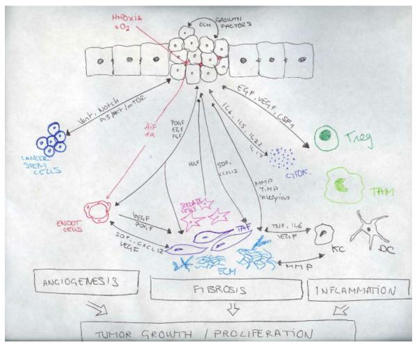
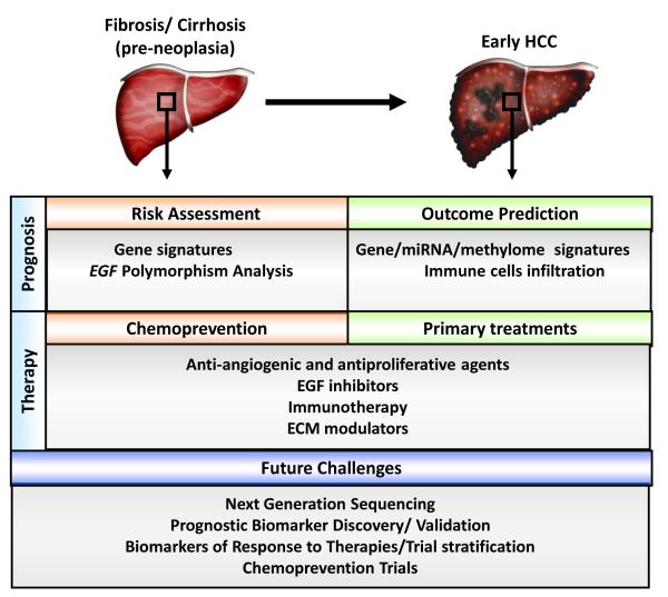
Hepatocellular carcinoma (HCC) is the most common primary liver tumor and the third greatest cause of cancer-related death worldwide, and its incidence is increasing. Despite the significant improvement in management of HCC over the past 30 years, there are no effective chemoprevention strategies, and only one systemic therapy has been approved for patients with advanced tumors. This drug, sorafenib, acts on tumor cells and the stroma. HCC develops from chronically damaged tissue that contains large amounts of inflammation and fibrosis, which also promote tumor progression and resistance to therapy. Increasing our understanding of how stromal components interact with cancer cells and the signaling pathways involved could help identify new therapeutic and chemopreventive targets.


Figures

Figure 1
Figure 1
Cellular components of the microenvironment and molecular mechanism influencing tumor growth and progression. Stromal, inflammatory and cancer cells interact among them to create a complex interaction network that origin a permissive microenvironment and favor tumor progression. TAFs, tumor associated fibroblasts; CSF-1, colony stimulating factor 1; EC, endothelial cells; KC, Kupffer cells; VEGF, vascular endothelial growth factor; FGF, Fibroblast growth factor PDGF, platelet-derived endothelial cell growth factor: Tregs, regulatory T cells; HGF, Hepatocyte Growth Factor; EGFR, Epidermal Growth Factor Receptor, MMPs, metaloproteinases; TIMP, Tissue Inhibitor of Metalloproteinases; HIF-1, HIF-1, hypoxia-inducible factor 1; TAM, Tumor associated macrophages; SDF-1, stromal cell-derived factor 1; CSC, Cancer stem cells; DC, Dendritic cells; TNF, Tumor Necrosis Factor.
Figure 1
Figure 1
Cellular components of the microenvironment and molecular mechanism influencing tumor growth and progression. Stromal, inflammatory and cancer cells interact among them to create a complex interaction network that origin a permissive microenvironment and favor tumor progression. TAFs, tumor associated fibroblasts; CSF-1, colony stimulating factor 1; EC, endothelial cells; KC, Kupffer cells; VEGF, vascular endothelial growth factor; FGF, Fibroblast growth factor PDGF, platelet-derived endothelial cell growth factor: Tregs, regulatory T cells; HGF, Hepatocyte Growth Factor; EGFR, Epidermal Growth Factor Receptor, MMPs, metaloproteinases; TIMP, Tissue Inhibitor of Metalloproteinases; HIF-1, HIF-1, hypoxia-inducible factor 1; TAM, Tumor associated macrophages; SDF-1, stromal cell-derived factor 1; CSC, Cancer stem cells; DC, Dendritic cells; TNF, Tumor Necrosis Factor.
Figure 2
Figure 2
Pathological features that may be present in hepatocellular carcinoma (HCC). (a) Poorly-differentiated HCC. Tumor cells have marked pleomorphic nuclei and an inflammatory infiltrate consisting of neutrophils. Ballooning degeneration and production of Mallory’s hyalines are also noted. H&E original magnification 400X. (b) Poorly-differentiated HCC with tumor cells arranged in a solid pattern. A focus of lymphocytic inflammatory infiltrate is present. H&E, original magnification 100X. (c) Well-differentiated multinodular HCC with dense fibrosis forming a wide septum that separate two HCC nodules. H&E, original magnification 40X. (d ) Increased vascularization in HCC. Vessels are highlighted by CD34 immunostaining. These vessels are nourishing the tumor. Original magnification 100X. Images courtesy of Dr. M. Isabel Fiel, Mount Sinai School of Medicine.
Figure 3
Figure 3
Anatomical and cellular alterations leading to HCC development. (a) Normal liver parenchyma. Hepatocytes with microvilli and fenestrated sinusoidal cells that favours the metabolic exchange. Space of Disse with few quiescence stellate cells containing lipid droplets. (b) Fibrotic liver. Upon chronic liver injury, hepatocytes loose the microvilli and sinusoids their fenestration, stellate cells become activated, loosing the lipid droplets and secreting ECM. (c) Hepaocellular carcinoma. Malignant transformation of hepatocytes with uncontrolled growth. Infiltration of inflammatory cells and cytokines. Development of new vessels (neoangiogenesis) and extense fibrosis with recruitment of tumor associated fibroblasts and cancer stem cells.
Figure 4
Figure 4
Schematic representation of therapeutic opportunities and application of prognostic biomarkers in the management of patients with HCC and pre-neoplastic conditions.

References

    1. Llovet JM, Burroughs A, Bruix J. Hepatocellular carcinoma. Lancet. 2003;362:1907–17. - PubMed
    1. Jemal A, Bray F, Center MM, et al. Global cancer statistics. CA Cancer J Clin. 61:69–90. - PubMed
    1. Villanueva A, Llovet JM. Targeted therapies for hepatocellular carcinoma. Gastroenterology. 140:1410–26. - PMC - PubMed
    1. Llovet JM, Ricci S, Mazzaferro V, et al. Sorafenib in advanced hepatocellular carcinoma. N Engl J Med. 2008;359:378–90. - PubMed
    1. Hanahan D, Coussens LM. Accessories to the crime: functions of cells recruited to the tumor microenvironment. Cancer Cell. 2012;21:309–22. - PubMed

Publication types

MeSH terms