. 2013 Mar;118(3):502-15.
doi: 10.1097/ALN.0b013e3182834d77.

Selective anesthesia-induced neuroinflammation in developing mouse brain and cognitive impairment

Affiliations

Selective anesthesia-induced neuroinflammation in developing mouse brain and cognitive impairment

Xia Shen et al. Anesthesiology. 2013 Mar.

Abstract

Background: : Recent population studies have suggested that children with multiple exposures to anesthesia and surgery at an early age are at an increased risk of cognitive impairment. The authors therefore have established an animal model with single versus multiple exposures of anesthetic(s) in young versus adult mice, aiming to distinguish the role of different types of anesthesia in cognitive impairment.

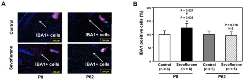
Methods: : Six- and 60-day-old mice were exposed to various anesthesia regimens. The authors then determined the effects of the anesthesia on learning and memory function, levels of proinflammatory cytokine interleukin-6 and tumor necrosis factor-α in brain tissues, and the amount of ionized calcium-binding adaptor molecule 1-positive cells, the marker of microglia activation, in the hippocampus.

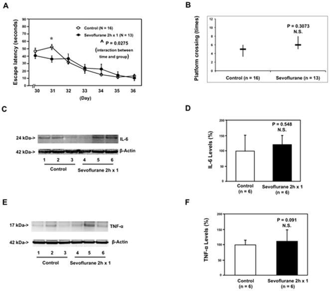
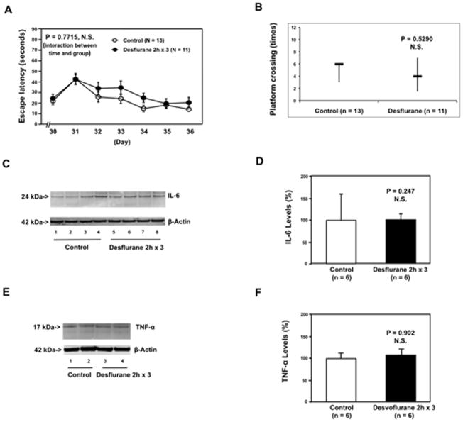
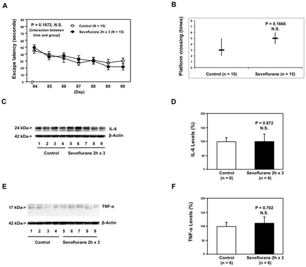
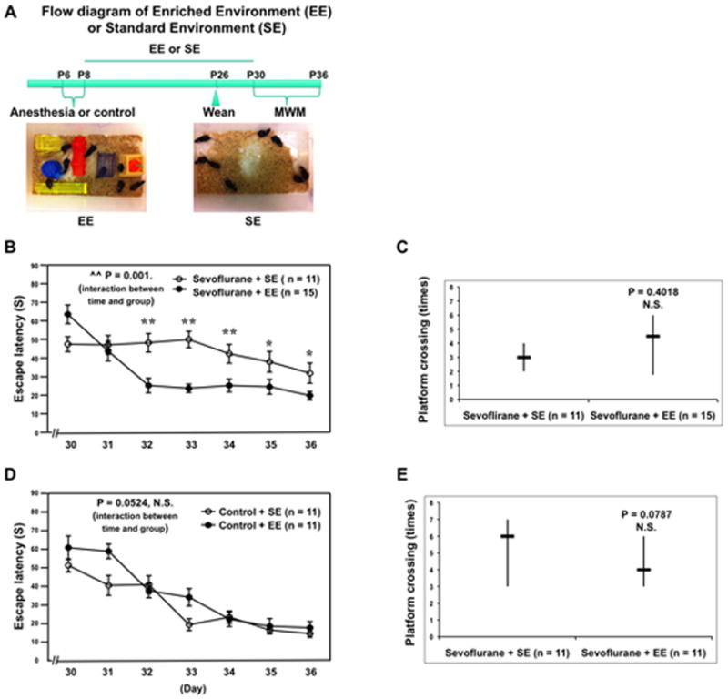
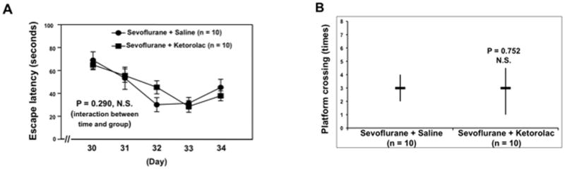
Results: : In this article, the authors show that anesthesia with 3% sevoflurane for 2 h daily for 3 days induced cognitive impairment and neuroinflammation (e.g., increased interleukin-6 levels, 151 ± 2.3% [mean ± SD] vs. 100 ± 9.0%, P = 0.035, n = 6) in young but not in adult mice. Anesthesia with 3% sevoflurane for 2 h daily for 1 day and 9% desflurane for 2 h daily for 3 days induced neither cognitive impairment nor neuroinflammation. Finally, an enriched environment and antiinflammatory treatment (ketorolac) ameliorated the sevoflurane-induced cognitive impairment.

Conclusions: : Anesthesia-induced cognitive impairment may depend on developmental stage, anesthetic agent, and number of exposures. These findings also suggest the cellular basis and the potential prevention and treatment strategies for anesthesia-induced cognitive impairment, which may ultimately lead to safer anesthesia care and better postoperative outcomes for children.


Figures

Figure 1
Figure 1. Anesthesia with 3% sevoflurane two hours daily for three days in P6 mice induces cognitive impairment in the mice tested from P30 to P36, and increases IL-6 and TNF-α levels in the brain tissues of the mice
A. Anesthesia with 3% sevoflurane two hours daily for three days in P6 mice increases the escape latency of mice swimming in the Morris Water Maze (MWM) as compared to the control condition (tested from P30 to P36) (control: n = 13, sevoflurane: n = 15). Two way ANOVA with repeated measurement analysis shows that there is a statistically significant interaction of time and group based on escape latency of MWM between mice following the control condition and mice following the sevoflurane anesthesia in the MWM. Specifically, mice that received the sevoflurane anesthesia had a longer escape latency time at P33, P34, P35 and P36 as compared to the control condition. B. Anesthesia with 3% sevoflurane two hours daily for three days in P6 mice reduces the platform crossing times of mice swimming in the MWM as compared to the control condition tested at P36 (control: n = 13, sevoflurane: n = 15). C. The sevoflurane anesthesia increases IL-6 levels in the brain tissues of the mice as compared to the control condition. There is no significant difference in β-Actin levels in the brain tissues of the mice between the sevoflurane anesthesia and control conditions. D. Quantification of the Western blot (n = 6) shows that the sevoflurane anesthesia increases IL-6 levels in the brain tissues of the mice as compared to the control condition. E. Sevoflurane anesthesia increases TNF-α levels in the brain tissues of the mice as compared to the control condition. There is no significant difference in β-Actin levels in brain tissues of the mice between the sevoflurane anesthesia and control groups. F. Quantification of the Western blot (n = 6) shows that the sevoflurane anesthesia increases TNF-α levels in the brain tissues of the mice as compared to the control group. P, postnatal day; MWM, Morris Water Maze; ANOVA, analysis of variance. IL, interleukin; TNF, tumor necrosis factor.
Figure 2
Figure 2. Anesthesia with 3% sevoflurane two hours daily for one day in P6 mice does not induce cognitive impairment in the mice tested from P30 to P36, and does not increase IL-6 and TNF-α levels in the brain tissues of the mice
A. Anesthesia with 3% sevoflurane two hours daily for one day in P6 mice does not increase escape latency of mice swimming in the MWM as compared to the control condition (tested from P30 to P36). Two way ANOVA with repeated measurement analysis shows that there is a statistically significant interaction of time and group based on escape latency of MWM between the mice following the control condition and mice following the sevoflurane anesthesia in the MWM (control: n = 16, sevoflurane: n = 13). Specifically, mice that received the sevoflurane anesthesia have a shorter escape latency time at P31 as compared to the control condition. B. Anesthesia with 3% sevoflurane two hours daily for one day in P6 mice does not reduce the platform crossing times of mice swimming in the MWM as compared to the control condition tested at P36 (control: n = 16, sevoflurane: n = 13). C. The sevoflurane anesthesia does not increase IL-6 levels in the brain tissues of the mice as compared to the control group. There is no significant difference in β-Actin levels in the brain tissues of the mice between the sevoflurane anesthesia and control groups. D. The quantification of the Western blot (n = 6) shows that the sevoflurane anesthesia does not increase IL-6 levels in the brain tissues of the mice as compared to the control group. E. The sevoflurane anesthesia does not increase TNF-α levels in the brain tissues of the mice as compared to the control condition. There is no significant difference in β-Actin levels in the brain tissues of the mice between the sevoflurane anesthesia and control conditions. F. The quantification of the Western blot (n = 6) shows that the sevoflurane anesthesia does not increase TNF-α levels in the brain tissues of the mice as compared to control condition. P, postnatal day; MWM, Morris Water Maze; ANOVA, analysis of variance. IL, interleukin; TNF, tumor necrosis factor.
Figure 3
Figure 3. Anesthesia with 9% desflurane two hours daily for three days in P6 mice does not induce cognitive impairment in the mice tested from P30 to P36, and does not increase IL-6 and TNF-α levels in the brain tissues of the mice
A. Anesthesia with 9% desflurane two hours daily for three days in P6 mice dose not increase escape latency of mice swimming in the MWM as compared to the control condition (tested from P30 to P36). Two way ANOVA with repeated measurement analysis shows that there is no statistically significant interaction of time and group based on escape latency of MWM between mice following the control condition and mice following the desflurane anesthesia in the MWM (control: n = 13, sevoflurane: n = 11). B. Anesthesia with 9% desflurane two hours daily for three days in P6 mice does not reduce the platform crossing times of mice swimming in the MWM as compared to the control condition tested at P36 (control: n = 13, sevoflurane: n = 11). C. The desflurane anesthesia does not increase IL-6 levels in the brain tissues of the mice as compared to the control condition. There is no significant difference in β-Actin levels in the brain tissues of the mice between the desflurane anesthesia and control groups. D. Quantification of the Western blot (n = 6) shows that the desflurane anesthesia does not increase IL-6 levels in the brain tissues of the mice as compared to the control condition. E. The desflurane anesthesia does not increase TNF-α levels in the brain tissues of the mice as compared to the control condition. There is no significant difference in β-Actin levels in the brain tissues of the mice between the desflurane anesthesia and control groups. F. Quantification of the Western blot (n = 6) shows that the desflurane anesthesia does not increase TNF-α levels in the brain tissues of the mice as compared to the control condition. P, postnatal day; MWM, Morris Water Maze; ANOVA, analysis of variance. IL, interleukin; TNF, tumor necrosis factor.
Figure 4
Figure 4. Anesthesia with 3% sevoflurane two hours daily for three days in P60 mice induce neither cognitive impairment in the mice tested from P84 to P90 nor increases in levels of IL-6 and TNF-α in the brain tissues of the mice
A. Anesthesia with 3% sevoflurane two hours daily for three days in P60 mice does not increase escape latency of mice swimming in the MWM as compared to the control condition (tested from P84 to P90). Two way ANOVA with repeated measurement analysis (control: n = 15, sevoflurane: n = 15) shows that there is no significant difference in the learning curve based on escape latency between mice following the control condition and mice following the sevoflurane anesthesia in the MWM. B. Anesthesia with 3% sevoflurane two hours daily for three days in P60 mice does not reduce the platform crossing times of mice swimming in the MWM as compared to the control condition tested at P90 (control: n = 15, sevoflurane: n = 15). C. The sevoflurane anesthesia does not increase IL-6 levels in the brain tissues of the mice as compared to the control condition. There is no significant difference in β-Actin levels in the brain tissues of the mice between the sevoflurane anesthesia and control groups. D. Quantification of the Western blot (n = 6) shows that the sevoflurane anesthesia does not increase IL-6 levels in the brain tissues of the mice as compared to the control condition. E. The sevoflurane anesthesia does not increase TNF-α levels in the brain tissues of the mice as compared to the control condition. There is no significant difference in β-Actin levels in the brain tissues of the mice between the sevoflurane anesthesia and control conditions. F. Quantification of the Western blot (n = 6) shows that the sevoflurane anesthesia does not increase TNF-α levels in the brain tissues of the mice as compared to the control group. IL, interleukin; TNF, tumor necrosis factor; P, postnatal day; MWM, Morris Water Maze; ANOVA, analysis of variance.
Figure 5
Figure 5. Anesthesia with 3% sevoflurane two hours daily for three days increases the amount of IBA1 positive cells in the hippocampus of P6, but not P60, mice
A. The sevoflurane anesthesia increases the amount of IBA1 positive cells in the hippocampus of P6 mice (the left column), but not P60 mice (right column), harvested at the end of the three-day anesthesia. B. Quantification of the immunohistochemistry image (n = 6) shows that the sevoflurane anesthesia increases the amount of IBA1 positive cells in the hippocampus of P6 mice (black bar versus white bar), but not in P60 mice (net bar versus gray bar). The two-way ANOVA shows that there is a significant interaction and that young age potentiates the sevoflurane anesthesia-induced increases in the amount of IBA1 positive cells. P, postnatal day; IBA1, ionized calcium binding adaptor molecule 1. ANOVA, analysis of variance.
Figure 6
Figure 6. EE attenuates the sevoflurane-induced cognitive impairment in mice
A. The flow diagram and pictures of EE and SE. B. Two-way ANOVA with repeated measurement analysis shows that there is a statistically significant interaction of time and group based on escape latency in MWM between mice following sevoflurane anesthesia plus SE and sevoflurane anesthesia plus EE (control: n = 11, sevoflurane: n = 15). C. The Mann-Whitney test shows that the platform crossing times of mice swimming in the MWM following the sevoflurane anesthesia plus EE is not significantly different from that of mice following the sevoflurane anesthesia plus SE (control: n = 11, sevoflurane: n = 15). D. Two-way ANOVA with repeated measurement analysis shows that there is no statistically significant interaction of time and group based on escape latency of MWM between the control condition plus SE and the control condition plus EE (control: n = 11, sevoflurane: n = 11). E. The Mann-Whitney test shows that there is no significant difference in platform crossing times of mice swimming in the MWM between the control condition plus SE and the control condition plus EE (control: n = 11, sevoflurane: n = 11). MWM, Morris Water Maze; ANOVA, analysis of variance; SE, standard environment; EE, enriched environment.
Figure 7
Figure 7. Ketorolac attenuates the sevoflurane-induced cognitive impairment in mice
A. Two-way ANOVA with repeated measurement analysis shows that there is no statistically significant interaction of time and group based on escape latency of MWM between the sevoflurane plus saline and sevoflurane plus ketorolac treated mice (control: n = 10, sevoflurane: n = 10). B. The Mann-Whitney test shows that there is no significant difference in platform crossing times of mice swimming in the MWM between the sevoflurane plus saline and sevoflurane plus ketorolac (control: n = 10, sevoflurane: n = 10). MWM, Morris Water Maze; ANOVA, analysis of variance.

References

    1. DeFrances CJ, Cullen KA, Kozak LJ. National Hospital Discharge Survey: 2005 annual summary with detailed diagnosis and procedure data. Vital Health Stat. 2007;13:1–209. - PubMed
    1. Rappaport B, Mellon RD, Simone A, Woodcock J. Defining safe use of anesthesia in children. N Engl J Med. 2011;364:1387–90. - PubMed
    1. Sun L. Early childhood general anaesthesia exposure and neurocognitive development. Br J Anaesth. 2010;105:61–8. - PMC - PubMed
    1. Wilder RT, Flick RP, Sprung J, Katusic SK, Barbaresi WJ, Mickelson C, Gleich SJ, Schroeder DR, Weaver AL, Warner DO. Early exposure to anesthesia and learning disabilities in a population-based birth cohort. Anesthesiology. 2009;110:796–804. - PMC - PubMed
    1. Flick RP, Katusic SK, Colligan RC, Wilder RT, Voigt RG, Olson MD, Sprung J, Weaver AL, Schroeder DR, Warner DO. Cognitive and behavioral outcomes after early exposure to anesthesia and surgery. Pediatrics. 2011;128:e1053–61. - PMC - PubMed

Publication types

MeSH terms