. 2013 Feb;20(2):230-236.
doi: 10.1038/nsmb.2485. Epub 2013 Jan 13.

Complexes of HIV-1 RT, NNRTI and RNA/DNA hybrid reveal a structure compatible with RNA degradation

Affiliations

Complexes of HIV-1 RT, NNRTI and RNA/DNA hybrid reveal a structure compatible with RNA degradation

Mikalai Lapkouski et al. Nat Struct Mol Biol. 2013 Feb.

Abstract

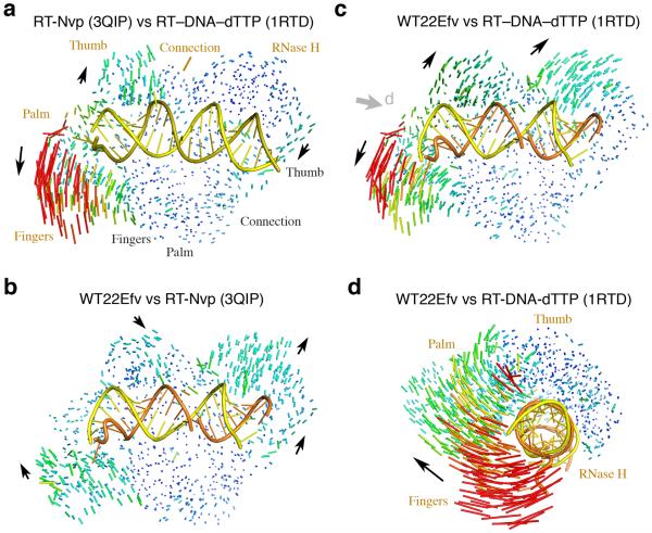
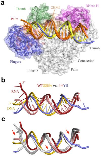
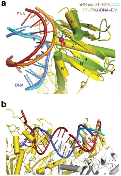
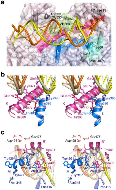
Hundreds of structures of type 1 human immunodeficiency virus (HIV-1) reverse transcriptase (RT) have been determined, but only one contains an RNA/DNA hybrid. Here we report three structures of HIV-1 RT complexed with a non-nucleotide RT inhibitor (NNRTI) and an RNA/DNA hybrid. In the presence of an NNRTI, the RNA/DNA structure differs from all prior nucleic acid-RT structures including the RNA/DNA hybrid. The enzyme structure also differs from all previous RT-DNA complexes. Thus, the hybrid has ready access to the RNase-H active site. These observations indicate that an RT-nucleic acid complex may adopt two structural states, one competent for DNA polymerization and the other for RNA degradation. RT mutations that confer drug resistance but are distant from the inhibitor-binding sites often map to the unique RT-hybrid interface that undergoes conformational changes between two catalytic states.


Figures

Figure 1
Figure 1
HIV-1 RT complexed with an RNA/DNA hybrid and an NNRTI. (a) The WT22Efv structure. RT is shown as molecular surface with p66 in wheat and p51 in silver. The polymerase and RNase H active site are highlighted in yellow and cyan, respectively. CN stands for connection domain. The hybrid is shown as tube-and-ladder with RNA in red and DNA in blue. The nucleic acid sequence is shown in the same color scheme above. The “x” denotes the abasic tetrahydrofuran substitution, and black letters indicate bases that are not traceable. The nick in the RNA strand is indicated by a black arrow. (b) The RNA (red), DNA (blue) and Efavirenz (bright orange) in the WT22Efv structure are shown with the simulated annealing omit Fo-Fc map contoured at 4.0 σ. The purple arrowheads in (a) and (b) mark the unique bend in the hybrid that allows it to reach the RNase H active site. (c) The ternary complex structure 1RTD representing all previously reported RT–DNA complexes. It is shown with dsDNA in light and dark blue and dTTP in green. The RT–DNA crosslinking site (Q258C) is marked. Four α-helices, αA (p66 fingers), αK (p66 CN), αL (p51 CN), and αM (p51), are shown as cylinders to highlight different RT structures in (a) and (c). A white arrowhead indicates the absence of the bend in the DNA. (d) Pairwise superposition of hybrids in the three RT complexes. Each structure is color-coded as labeled.
Figure 2
Figure 2
Structure comparison of HIV-1 RT. (a) An RT–DNA–dTTP ternary complex (1RTD) versus an RT–NNRTI (3QIP) complex. Differences are shown as a pairwise Cα-Cα vector map after superposition of p51. Increase in vector length is shown in blue (0.3 Å) to red (>8Å). The DNA duplex in 1RTD is shown as yellow and olive tube-and-ladder. (b) The WT22Efv structure versus 3QIP. The RNA (orange) and DNA (yellow) hybrid is also shown. Black arrows indicate the direction of movement from 3QIP to WT22Efv structure. (c) Comparison of WT22Efv with 1RTD after superposition of p51 as in (b). (d) Superposition of thumb, connection and RNase H in p66 between WT22Efv and 1RTD. The viewing angle is indicated by the grey arrowhead in panel c.
Figure 3
Figure 3
Structural comparison of WT22Efv and RT–DNA–Nvp (3V81). (a) Pairwise comparisons of RT in the 3V81 structure with RT complexed with Nevirapine (3QIP), an RT ternary complex (1RTD), and our WT22Efv (4B3O). The Cα-Cα vector maps are shown after superposition of p51 subunits and colored as in Fig. 2. Domain movements of 3V81 relative to others are indicated by black arrowheads. (b) Comparison of the entire RT–DNA–Nvp complex (3V81) with an RT–DNA–dNTP ternary complex (3KK2). Upon superposition of the nucleic acids, both of which are crosslinked to p66 via Q258C, not only are the DNAs highly similar, but also are the RT proteins except for the p66 fingers and palm subdomains as indicated by the grey arrowhead. The protein and DNA are color coded as indicated, and the NNRTI (Nvp) is shown as purple spheres.
Figure 4
Figure 4
Structural comparison of nucleic acid complexed with RT. (a) Five previously reported RT–nucleic acid complexes (1RTD, 1HYS, 1T05, 2HMI (uncrosslinked), and 3KJV). The structures are shown after superposition of the p51 subunits. RT is shown as molecular surface with the fingers, palm, thumb, connection and RNase H domains color-coded blue, red, green, gold and magenta, respectively. The four DNA are depicted as orange template and yellow primer strands. The PPT RNA/DNA hybrid in 1HYS is shown in pink and blue. (b) Differences between the RNA/DNA hybrid in WT22Efv (red and yellow) and the PPT hybrid in 1HYS (pink and blue) after superposition of p51 subunits. (c) Comparison of the two hybrids and four DNAs (in light grey as in (a)). Red arrows highlight the main changes in WT22Efv.
Figure 5
Figure 5
The RNA/DNA hybrid structure is compatible with RNase H cleavage. (a) Comparison of WT22Efv with human RNase H1–substrate complex (2QK9). Human RNase H1 (green) is superimposed with the RNase H domain of HIV-1 RT (gold). The two hybrids are colored orange and cyan (human) and red and blue (WT22Efv). The RT active site is marked by the conserved carboxylates (red sticks) and the cation (purple sphere) in the hRNase H1 structure. Protein secondary structures are labeled according to convention. (b) Compatibility with RNA degradation. Nucleic acids in the WT22Efv and human RNase H1 structures can be readily linked after superposition of the RNase H domain. p51 and p66 are depicted in silver and gold, respectively.
Figure 6
Figure 6
The altered p51–p66 subunit interface interacts with the RNA/DNA hybrid. (a) Overview of the WT22Efv structure with the altered subunit interface highlighted in blue (p51) and pink (p66). NNRTI and NRTI resistance mutations are found in the p66 connection and RNase H domain (pea green) that contacts the widened major groove. Residues surrounding Asn348 in p51 and forming van der Waals contacts with the RNA strand are colored teal. (b) A close-up stereo view of the interfacial helices between the p51 (blue) and p66 (pink) connection domains. The altered protein interface relative to 1RTD (an RT–DNA ternary complex, in lighter shades) is correlated with the change from DNA binding (pale yellow and wheat) to RNA/DNA hybrid (orange/yellow) binding. (c) A close-up stereo view of the back of (a). This shows the p51 C-terminus (blue) and helix αB′ (pink) in the WT22Efv structure and in 3KK2 (an RT–DNA ternary complex complete with helix αM in p51, in lighter shades). The hybrid in the WT22Efv (orange and yellow) and DNA in 3KK2 are also shown. In panels (b) and (c), the p51 subunits are superimposed.

Comment in

References

    1. Varmus H. Reverse transcription. Sci Am. 1987;257:56–59. 62–54. - PubMed
    1. Sarafianos SG, et al. Structure and function of HIV-1 reverse transcriptase: molecular mechanisms of polymerization and inhibition. J Mol Biol. 2009;385:693–713. - PMC - PubMed
    1. Cihlar T, Ray AS. Nucleoside and nucleotide HIV reverse transcriptase inhibitors: 25 years after zidovudine. Antiviral Res. 2010;85:39–58. - PubMed
    1. de Bethune MP. Non-nucleoside reverse transcriptase inhibitors (NNRTIs), their discovery, development, and use in the treatment of HIV-1 infection: a review of the last 20 years (1989–2009) Antiviral Res. 2010;85:75–90. - PubMed
    1. di Marzo Veronese F, et al. Characterization of highly immunogenic p66/p51 as the reverse transcriptase of HTLV-III/LAV. Science. 1986;231:1289–1291. - PubMed

Publication types

MeSH terms

LinkOut - more resources