

Behavior of the P1.HTR mastocytoma cell line implanted in the chorioallantoic membrane of chick embryos
- PMID: 23314344
- PMCID: PMC3854347
- DOI: 10.1590/1414-431x20122434
Behavior of the P1.HTR mastocytoma cell line implanted in the chorioallantoic membrane of chick embryos
Abstract
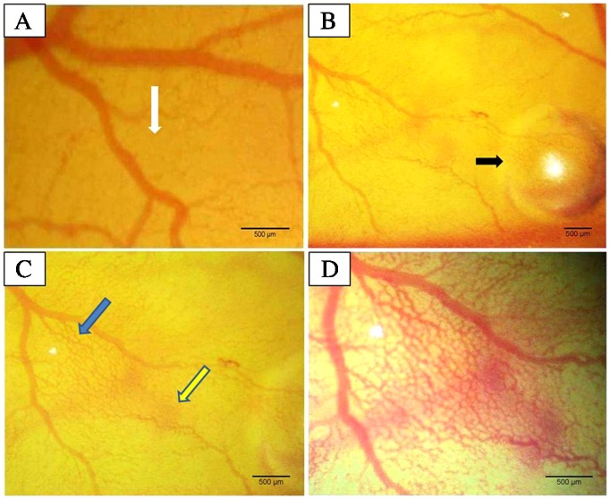
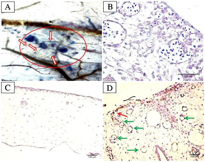
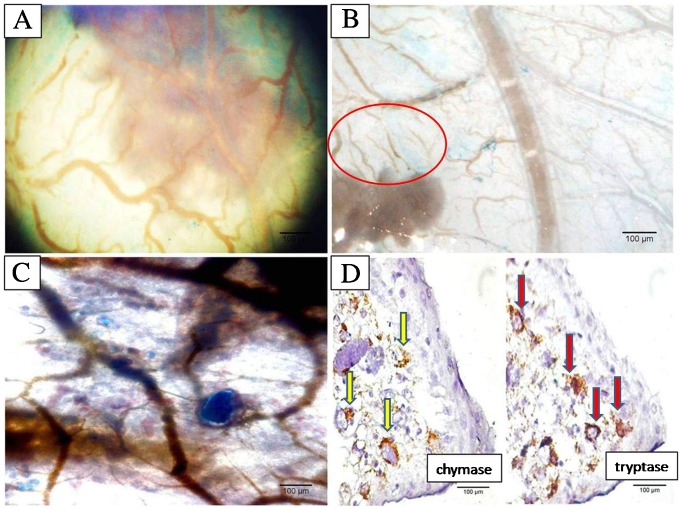
The P1.HTR cell line includes highly transfectable cells derived from P815 mastocytoma cells originating from mouse breast tissue. Despite its widespread use in immunogenic studies, no data are available about the behavior of P1.HTR cells in the chick embryo chorioallantoic membrane model. The objective of the present investigation was to study the effects of P1.HTR cells implanted on the chorioallantoic membrane of chick embryos. We inoculated P1.HTR cells into the previously prepared chick embryo chorioallantoic membrane and observed the early and late effects of these cells by stereomicroscopy, histochemistry and immunohistochemistry. A highly angiotropic and angiogenic effect occurred early after inoculation and a tumorigenic potential with the development of mastocytoma keeping well mast cells immunophenotype was detected later during the development. The P1.HTR mastocytoma cell line is a good tool for the development of the chick embryo chorioallantoic membrane mastocytoma model and also for other studies concerning the involvement of blood vessels. The chick embryo chorioallantoic membrane model of mastocytoma retains the mast cell immunophenotype under experimental conditions and could be used as an experimental tool for in vivo preliminary testing of antitumor and antivascular drugs.
Figures
Similar articles
-
Protocol for performing angiogenic and tumorigenic assays using the in ovo chick embryo chorioallantoic membrane model.STAR Protoc. 2025 Mar 21;6(1):103663. doi: 10.1016/j.xpro.2025.103663. Epub 2025 Feb 28. STAR Protoc. 2025. PMID: 40022735 Free PMC article.
-
Methods for Analyzing Tumor Angiogenesis in the Chick Chorioallantoic Membrane Model.Methods Mol Biol. 2016;1406:255-69. doi: 10.1007/978-1-4939-3444-7_22. Methods Mol Biol. 2016. PMID: 26820962
-
The chick embryo chorioallantoic membrane as a model to study tumor metastasis.Angiogenesis. 2008;11(4):311-9. doi: 10.1007/s10456-008-9117-1. Epub 2008 Sep 9. Angiogenesis. 2008. PMID: 18780151 Review.
-
The chick embryo chorioallantoic membrane as a model for tumor biology.Exp Cell Res. 2014 Nov 1;328(2):314-24. doi: 10.1016/j.yexcr.2014.06.010. Epub 2014 Jun 24. Exp Cell Res. 2014. PMID: 24972385 Review.
-
The chick embryo chorioallantoic membrane as an in vivo experimental model to study multiple myeloma.Enzymes. 2019;46:23-35. doi: 10.1016/bs.enz.2019.08.006. Epub 2019 Oct 18. Enzymes. 2019. PMID: 31727275 Review.
Cited by
-
Antiangiogenic Potential of Beneficial Sterols from Parotoid Gland Secretion of Indian Common Toads (Duttaphrynus melanostictus) in the Coastal Region of the Indian Subcontinent: An In Vivo to In Silico Approach.ACS Omega. 2025 Mar 4;10(10):10480-10492. doi: 10.1021/acsomega.4c10809. eCollection 2025 Mar 18. ACS Omega. 2025. PMID: 40124047 Free PMC article.
References
-
- Metcalfe DD, Baram D, Mekori YA. Mast cells. Physiol Rev. 1997;77:1033–1079. - PubMed
-
- Crivellato E, Beltrami CA, Mallardi F, Ribatti D. The mast cell: an active participant or an innocent bystander? Histol Histopathol. 2004;19:259–270. - PubMed
-
- Duse AO, Ceausu RA, Mezei T, Cimpean AM, Gaje P, Ionita H, et al. Mast cells contribute to the angiogenesis in non-Hodgkin lymphoma. An immunohistochemical study based on the relationship with microvessel density. Rom J Morphol Embryol. 2011;52:1091–1096. - PubMed
-
- Liu J, Zhang Y, Zhao J, Yang Z, Li D, Katirai F, et al. Mast cell: insight into remodeling a tumor microenvironment. Cancer Metastasis Rev. 2011;30:177–184. doi:10.1007/s10555-011-9276-1. - PubMed
Publication types
MeSH terms
LinkOut - more resources
Full Text Sources
Other Literature Sources
