

Nerve growth factor reduces myocardial ischemia/reperfusion injury in rat hearts
- PMID: 23314533
- PMCID: PMC3938895
- DOI: 10.1515/jbcpp-2012-0045
Nerve growth factor reduces myocardial ischemia/reperfusion injury in rat hearts
Abstract
Background: Nerve growth factor (NGF) is a neurotrophin that supports the survival and differentiation of sympathetic neurons, and its increased expression after myocardial infarct was correlated with cardiac sympathetic hyperinnervation and arrhythmias. However, it is unclear whether NGF protects the heart during infarct. In this study, we sought to address this issue in rat heart exposed to ischemia/reperfusion injury (IRI).
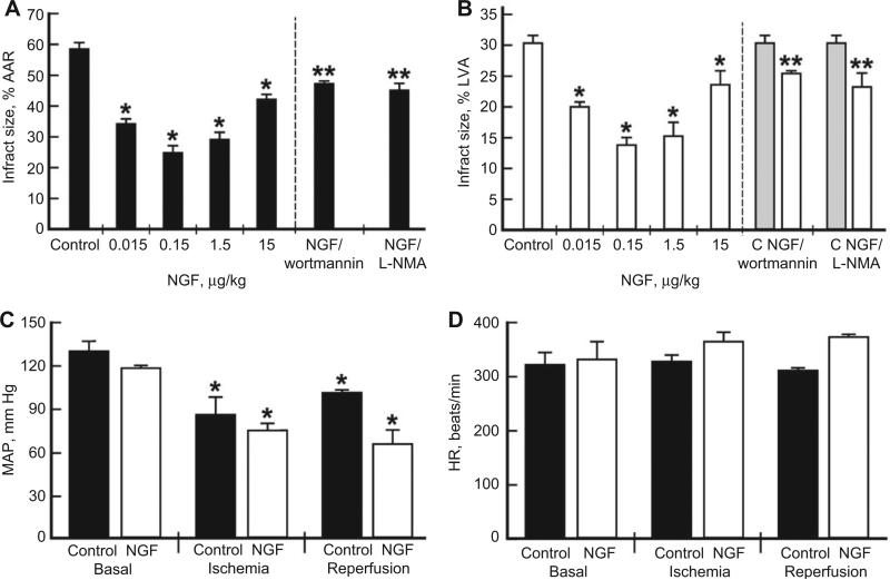
Methods: NGF was administered intravenously (IV), 15 min before ischemia, at different concentrations in the absence or presence of inhibitors of phosphatidylinositol-3 kinase (PI3K) or nitric oxide synthase (NOS) in different groups of rats (n=6) with left coronary occlusion for 30 min followed by 120-min reperfusion. The area at risk and infarct to risk ratios were determined from sections stained with 1% 2,3,5-triphenylterazolium chloride.
Results: NGF treatment at doses of 0.015-15 μg/kg, with an optimal dose of 0.15 μg/kg given IV before ischemia, reduced the infarct size from about 60% at the area of risk to about 25%, indicating cardioprotection by about 60%. The infarct-sparing effects of NGF were partially abolished by the inhibition of PI3K and NOS using wortmannin and N(G)-monomethyl-l-arginine, respectively.
Conclusions: We have demonstrated for the first time that NGF attenuates myocardial infarct damage in an in vivo rat model of myocardial regional IRI. This cardioprotective effect is proposed to be related to the activities of PI3K and NOS. This suggests that NGF has a potential therapeutic role in the treatment of IRI.
Figures
Similar articles
-
Nerve growth factor protects the ischemic heart via attenuation of the endoplasmic reticulum stress induced apoptosis by activation of phosphatidylinositol 3-kinase.Int J Med Sci. 2015 Jan 1;12(1):83-91. doi: 10.7150/ijms.10101. eCollection 2015. Int J Med Sci. 2015. PMID: 25552923 Free PMC article.
-
SCH 79797, a selective PAR1 antagonist, limits myocardial ischemia/reperfusion injury in rat hearts.Basic Res Cardiol. 2007 Jul;102(4):350-8. doi: 10.1007/s00395-007-0653-4. Epub 2007 Apr 30. Basic Res Cardiol. 2007. PMID: 17468933 Free PMC article.
-
Inhibition of Rho kinase protects from ischaemia-reperfusion injury via regulation of arginase activity and nitric oxide synthase in type 1 diabetes.Diab Vasc Dis Res. 2017 May;14(3):236-245. doi: 10.1177/1479164116687935. Epub 2017 Feb 9. Diab Vasc Dis Res. 2017. PMID: 28183205
-
PPAR-alpha activation protects the type 2 diabetic myocardium against ischemia-reperfusion injury: involvement of the PI3-Kinase/Akt and NO pathway.Am J Physiol Heart Circ Physiol. 2009 Mar;296(3):H719-27. doi: 10.1152/ajpheart.00394.2008. Epub 2009 Jan 16. Am J Physiol Heart Circ Physiol. 2009. PMID: 19151258
-
Reduced susceptibility to ischemia-induced arrhythmias in the preconditioned rat heart is independent of PI3-kinase/Akt.Physiol Res. 2009;58(3):443-447. doi: 10.33549/physiolres.931743. Physiol Res. 2009. PMID: 19627174
Cited by
-
Effects of Yiqi Huoxue Decoction on Post-Myocardial Infarction Cardiac Nerve Remodeling and Cardiomyocyte Hypertrophy in Rats.Evid Based Complement Alternat Med. 2021 Aug 21;2021:5168574. doi: 10.1155/2021/5168574. eCollection 2021. Evid Based Complement Alternat Med. 2021. PMID: 34471416 Free PMC article.
-
Nerve growth factor protects the ischemic heart via attenuation of the endoplasmic reticulum stress induced apoptosis by activation of phosphatidylinositol 3-kinase.Int J Med Sci. 2015 Jan 1;12(1):83-91. doi: 10.7150/ijms.10101. eCollection 2015. Int J Med Sci. 2015. PMID: 25552923 Free PMC article.
-
Nerve growth factor: a neuroimmune crosstalk mediator for all seasons.Immunology. 2017 May;151(1):1-15. doi: 10.1111/imm.12717. Epub 2017 Feb 21. Immunology. 2017. PMID: 28112808 Free PMC article. Review.
-
Nerve growth factor-induced Akt/mTOR activation protects the ischemic heart via restoring autophagic flux and attenuating ubiquitinated protein accumulation.Oncotarget. 2017 Jan 17;8(3):5400-5413. doi: 10.18632/oncotarget.14284. Oncotarget. 2017. PMID: 28036273 Free PMC article.
-
Pleiotropic activity of nerve growth factor in regulating cardiac functions and counteracting pathogenesis.ESC Heart Fail. 2021 Apr;8(2):974-987. doi: 10.1002/ehf2.13138. Epub 2021 Jan 19. ESC Heart Fail. 2021. PMID: 33465292 Free PMC article. Review.
References
-
- Levi-Montalcini R. The nerve growth factor 35 years later. Science. 1987;237:1154–62. - PubMed
-
- Lazarovici P, Marcinkiewicz C, Lelkes PI. Cross talk between the cardiovascular and nervous systems: neurotrophic effects of vascular endothelial growth factor (VEGF) and angiogenic effects of nerve growth factor (NGF) – implications in drug development. Curr Pharm Des. 2006;12:2609–22. - PubMed
-
- Govoni S, Pascale A, Amadio M, Calvillo L, D'Elia E, Cereda C, et al. NGF and heart: is there a role in heart disease ? Pharmacol Res. 2011;63:266–77. - PubMed
-
- Routhu KV, Tsopanoglou NE, Strande JL. Parstatin(1–26): the putative signal peptide of protease-activated receptor 1 confers potent protection from myocardial ischemia-reperfusion injury. J Pharmacol Exp Ther. 2010;332:898–905. - PubMed
Publication types
MeSH terms
Substances
Grants and funding
LinkOut - more resources
Full Text Sources
Other Literature Sources