

Ultrasonography of peripheral nerves
- PMID: 23314937
- PMCID: PMC3675797
- DOI: 10.1007/s11910-012-0328-x
Ultrasonography of peripheral nerves
Abstract
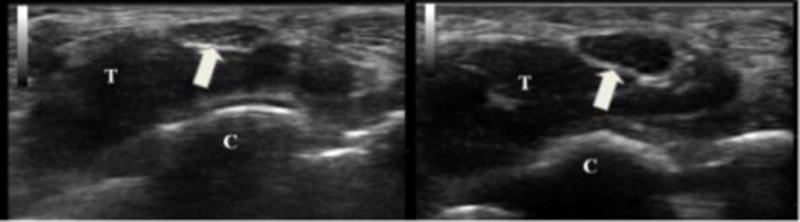
Over the last decade, neuromuscular ultrasonography has emerged as a useful tool for the diagnosis of peripheral nerve disorders. This article reviews sonographic findings of normal nerves, including key quantitative ultrasound measurements that are helpful in the evaluation of focal and possibly generalized peripheral neuropathies. It also discusses several recent articles outlining the evidence base for the use of this technology, as well as new findings in compressive, traumatic, and generalized neuropathies. Ultrasonography is well suited for use in electrodiagnostic laboratories, where physicians, experienced in both the clinical evaluation of patients and the application of hands-on technology, can integrate findings from the patient's history, physical examination, electrophysiological studies, and imaging for diagnosis and management.
Conflict of interest statement
No potential conflicts of interest relevant to this article were reported.
Figures
References
-
- Heckmatt JZ, Dubowitz V, Leeman S. Detection of pathological change in dystrophic muscle with B-scan ultrasound imaging. Lancet. 1980;1:1389–1390. - PubMed
-
- Fornage BD. Peripheral nerves of the extremities: imaging with US. Radiology. 1988;167:179–182. - PubMed
-
- Buchberger W, Schön G, Strasser K, Jungwirth W. High-resolution ultrasonography of the carpal tunnel. J Ultrasound Med. 1991;10:531–537. - PubMed
-
- Hullander M, Spillane W, Leivers D, Balsara Z. The use of Doppler ultrasound to assist with sciatic nerve blocks. Reg Anesth. 1991;16:282–284. - PubMed
-
- Nakamichi K, Tachibana S. Transverse sliding of the median nerve beneath the flexor retinaculum. J Hand Surg Br. 1992;17:213–216. - PubMed
Publication types
MeSH terms
Grants and funding
LinkOut - more resources
Full Text Sources
Other Literature Sources
Medical
