

IL-2R signaling is essential for functional maturation of regulatory T cells during thymic development
- PMID: 23315074
- PMCID: PMC3563871
- DOI: 10.4049/jimmunol.1201218
IL-2R signaling is essential for functional maturation of regulatory T cells during thymic development
Abstract
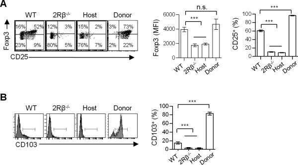
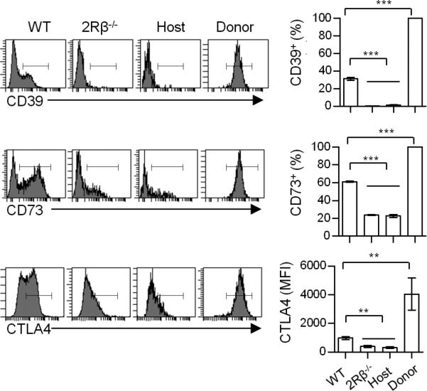
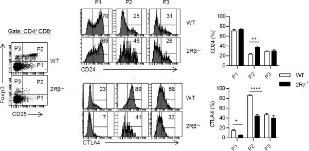
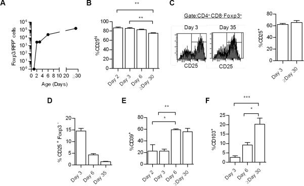
CD4(+) Foxp3(+) regulatory T cells (Tregs) are an independent cell lineage, and their developmental progression during thymic development depends on IL-2R signaling. However, the role of IL-2R signaling during thymic Treg development remains only partially understood. The current study assessed the contribution of IL-2 to the expansion and functional programming of developing Tregs. In the absence of IL-2Rβ signaling, predominantly CD4(+) CD25(-) Foxp3(lo) T cells were found, and these cells exhibited somewhat lower expression of the proliferative marker Ki67. These immature Tregs, which represent products of failed development, were also found in normal mice and were characterized by markedly lower expression of several Treg functional molecules. Therefore, IL-2R is required for the progression, functional programming, and expansion of Tregs during thymic development. An IL-2R-signaling mutant that lowers STAT5 activation readily supported Treg functional programming, but Treg proliferation remained somewhat impaired. The requirement for IL-2 during thymic Treg expansion was best illustrated in mixed chimeras where the Tregs with mutant IL-2Rs were forced to compete with wild-type Tregs during their development. Tregs with impaired IL-2R signaling were more prevalent in the thymus than spleen in these competitive experiments. The general effectiveness of mutant IL-2Rs to support thymic Treg development is partially accounted for by a heightened capacity of thymic Tregs to respond to IL-2. Overall, our data support a model in which limiting IL-2R signaling is amplified by thymic Tregs to readily support their development and functional programming, whereas these same conditions are not sufficient to support peripheral Treg homeostasis.
Figures


Similar articles
-
Essential and non-overlapping IL-2Rα-dependent processes for thymic development and peripheral homeostasis of regulatory T cells.Nat Commun. 2019 Mar 4;10(1):1037. doi: 10.1038/s41467-019-08960-1. Nat Commun. 2019. PMID: 30833563 Free PMC article.
-
A function for IL-7R for CD4+CD25+Foxp3+ T regulatory cells.J Immunol. 2008 Jul 1;181(1):225-34. doi: 10.4049/jimmunol.181.1.225. J Immunol. 2008. PMID: 18566388 Free PMC article.
-
IL-2Rbeta links IL-2R signaling with Foxp3 expression.Eur J Immunol. 2007 Jul;37(7):1817-26. doi: 10.1002/eji.200737101. Eur J Immunol. 2007. PMID: 17559173
-
The Molecular Control of Regulatory T Cell Induction.Prog Mol Biol Transl Sci. 2015;136:69-97. doi: 10.1016/bs.pmbts.2015.09.001. Epub 2015 Oct 9. Prog Mol Biol Transl Sci. 2015. PMID: 26615093 Review.
-
T-cell tolerance and the multi-functional role of IL-2R signaling in T-regulatory cells.Immunol Rev. 2011 May;241(1):63-76. doi: 10.1111/j.1600-065X.2011.01004.x. Immunol Rev. 2011. PMID: 21488890 Free PMC article. Review.
Cited by
-
Cell penetrating recombinant Foxp3 protein enhances Treg function and ameliorates arthritis.Biochem Biophys Res Commun. 2013 May 3;434(2):263-7. doi: 10.1016/j.bbrc.2013.02.114. Epub 2013 Mar 26. Biochem Biophys Res Commun. 2013. PMID: 23541572 Free PMC article.
-
[Expression of interleukin-9 in colon cancer tissues and its clinical significance].Nan Fang Yi Ke Da Xue Xue Bao. 2018 Jul 30;38(8):943-948. doi: 10.3969/j.issn.1673-4254.2018.08.07. Nan Fang Yi Ke Da Xue Xue Bao. 2018. PMID: 30187869 Free PMC article. Chinese.
-
IL2 is required for functional maturation of regulatory T cells.Anim Cells Syst (Seoul). 2016 Dec 30;21(1):1-9. doi: 10.1080/19768354.2016.1272489. eCollection 2017. Anim Cells Syst (Seoul). 2016. PMID: 30460045 Free PMC article.
-
Gfi1: a unique controller of T(reg) cells.Cell Cycle. 2013 Dec 1;12(23):3581-2. doi: 10.4161/cc.26824. Epub 2013 Oct 21. Cell Cycle. 2013. PMID: 24131921 Free PMC article. No abstract available.
-
Abnormal expression pattern of the IL-2 receptor β-chain on CD4+ T cells in ANCA-associated vasculitis.Dis Markers. 2014;2014:249846. doi: 10.1155/2014/249846. Epub 2014 Feb 9. Dis Markers. 2014. PMID: 24648606 Free PMC article.
References
-
- Malek TR, Bayer AL. Tolerance, not immunity, crucially depends on IL-2. Nat. Rev. Immunol. 2004;4:665–674. - PubMed
-
- Bayer AL, Yu A, Malek TR. Function of the IL-2R for thymic and peripheral CD4+CD25+ Foxp3+ T regulatory cells. J. Immunol. 2007;178:4062–4071. - PubMed
-
- Malek TR. The biology of interleukin-2. Annu Rev Immunol. 2008;26:453–479. - PubMed
-
- Fontenot JD, Rasmussen JP, Gavin MA, Rudensky AY. A function for interleukin 2 in Foxp3-expressing regulatory T cells. Nat. Immunol. 2005;6:1142–1151. - PubMed
Publication types
MeSH terms
Substances
Grants and funding
LinkOut - more resources
Full Text Sources
Other Literature Sources
Molecular Biology Databases
Research Materials
Miscellaneous
