. 2013 Feb 15;190(4):1672-84.
doi: 10.4049/jimmunol.1102462. Epub 2013 Jan 11.

EBV BILF1 evolved to downregulate cell surface display of a wide range of HLA class I molecules through their cytoplasmic tail

Affiliations

EBV BILF1 evolved to downregulate cell surface display of a wide range of HLA class I molecules through their cytoplasmic tail

Bryan D Griffin et al. J Immunol. .

Abstract

Coevolution of herpesviruses and their hosts has driven the development of both host antiviral mechanisms to detect and eliminate infected cells and viral ploys to escape immune surveillance. Among the immune-evasion strategies used by the lymphocryptovirus (γ(1)-herpesvirus) EBV is the downregulation of surface HLA class I expression by the virally encoded G protein-coupled receptor BILF1, thereby impeding presentation of viral Ags and cytotoxic T cell recognition of the infected cell. In this study, we show EBV BILF1 to be expressed early in the viral lytic cycle. BILF1 targets a broad range of HLA class I molecules, including multiple HLA-A and -B types and HLA-E. In contrast, HLA-C was only marginally affected. We advance the mechanistic understanding of the process by showing that the cytoplasmic C-terminal tail of EBV BILF1 is required for reducing surface HLA class I expression. Susceptibility to BILF1-mediated downregulation, in turn, is conferred by specific residues in the intracellular tail of the HLA class I H chain. Finally, we explore the evolution of BILF1 within the lymphocryptovirus genus. Although the homolog of BILF1 encoded by the lymphocryptovirus infecting Old World rhesus primates shares the ability of EBV to downregulate cell surface HLA class I expression, this function is not possessed by New World marmoset lymphocryptovirus BILF1. Therefore, this study furthers our knowledge of the evolution of immunoevasive functions by the lymphocryptovirus genus of herpesviruses.


Figures

Figure 1
Figure 1. The BILF1 gene is expressed early in the EBV lytic cycle
EBV lytic cycle gene expression was induced in AKBM cells by cross-linking of the B cell receptor with α-IgG for the indicated time periods (hours) (A), or for 16 h in the presence or absence of PAA (B). RNA was isolated, cDNA generated, and expression levels of the indicated gene products determined by PCR and EtBr gel electrophoresis.
Figure 2
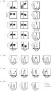
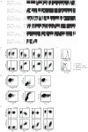
Figure 2. EBV BILF1 reduces surface expression of a broad range of HLA class I alleles
A, MJS cells were transduced with GFP-encoding (control) or EBV BILF1/GFP-encoding (BILF1) replication-deficient lentivirus. After 7 days, surface expression of FLAG-tagged EBV BILF1, total HLA class I, HLA class II, HLA-A1 and HLA-B8 was determined by flow cytometry. Transduced and untransduced MJS cells were mixed before antibody staining to allow comparison in a single assay (left and middle columns). Surface levels of the indicated proteins were also compared between GFP+ control cells and BILF1/GFP+ cells (histograms, right column). B, HeLa; C, 293T and D, U373 cells were transduced with GFP-encoding (control) or EBV BILF1/GFP-encoding (BILF1) lentivirus. Surface expression of FLAG-tagged EBV BILF1, total HLA class I or of the indicated HLA class I alleles was examined by flow cytometry after 7 days.
Figure 3
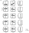
Figure 3. EBV BILF1 only marginally downregulates cell surface HLA-C expression
U937 cells were transduced with GFP-encoding (control) or EBV BILF1/GFP-encoding (BILF1) replication-deficient retrovirus. Surface expression of HA-tagged EBV BILF1, total HLA class I, HLA-A3 and HLA-B18 and HLA-C was determined by flow cytometry. Surface levels of the indicated proteins were compared between BILF1/GFP cells and BILF1+/GFP+ cells (histograms, right column).
Figure 4
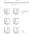
Figure 4. The EBV BILF1 C-terminal tail is required for HLA class I downregulation
A, Schematic representation of the 7-transmembrane EBV BILF1 vGPCR with the sequence of the 26 most C-terminally located amino acid residues described. The arrow indicates the point of truncation used to generate EBV BILF1-ΔC19. Dashed-line boxes indicate putative endocytosis/sorting motifs. B, EBV BILF1-ΔC19 activates intracellular signalling pathways. 293-NFκB-luciferase cells were co-transfected with control expression vector or constructs encoding EBV BILF1wt, EBV BILF1-K122A or EBV BILF1-ΔC19, and pGL3-Renilla (for normalizing transfection efficiency) luciferase. At 24 h post-transfection, cell lysates were assayed for firefly and Renilla luciferase activity. Data are presented as fold NFκB induction relative to cells transfected with vector alone. Results represent mean +/− SEM of a representative experiment performed in triplicate. C, MJS cells were transduced with GFP-encoding (control), EBV BILF1wt/GFP-encoding (BILF1wt) or EBV BILF1-ΔC19/GFP-encoding (ΔC19) replication-deficient lentivirus. After 7 days, surface expression of FLAG-tagged EBV BILF1, HLA-B8 and HLA class II was determined by flow cytometry.
Figure 5
Figure 5. The cytoplasmic region of HLA class I HC is required for BILF1-mediated cell surface downregulation
A, Sequence of the HLA-B8 C-terminus. The arrow denotes the point of truncation used to generate HLA-B8 short. B, U373 cells were transduced with HLA-B8wt/trNGFR-encoding or HLA-B8 short/trNGFR-encoding replication-deficient lentivirus. Surface expression of HLA-B8 and trNGFR on transduced and untransduced cells was determined by flow cytometry. C, U373-HLA-B8wt and U373-HLA-B8 short cells were further transduced with GFP-encoding (control) or EBV BILF1/GFP-encoding (BILF1) replication-deficient lentivirus. After 7 days, surface expression of HLA-B8 and the endogenous HLA-B18 allele was measured by flow cytometry.
Figure 6
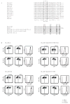
Figure 6. EBV BILF1-mediated HLA-B8 downregulation requires amino acid residues in the cytoplasmic region of the HLA-B8 molecule
A, upper panel, Amino acid sequence alignment of selected HLA class I heavy chain C-terminal tails. Alignments were generated using ClustalW2. An asterisk (*) indicates positions that have a single, fully conserved residue. A colon (:) indicates conservation between groups of strongly similar properties. A full stop (.) indicates conservation between groups of weakly similar properties. Sequences of the following subtypes were selected from the IMGT/HLA Database: A*01:01:01:01 (HLA-A1), A*02:01:01:01 (HLA-A2), A*03:01:01:01 (HLA-A3), A*68:01:01:01 (HLA-A68), B*07:02:01 (HLA-B7), B*08:01:01 (HLA-B8), B*15:03:01 (HLA-B15), B*18:01:01:01 (HLA-B18), C*01:02:01 (HLA-C), E*01:01:01:01 (HLA-E). The dashed box indicates a putative YXXA internalization motif present in HLA-A, -B and -E alleles but not HLA-C. A, lower panel, C-terminal tail amino acid sequences of HLA-B8 point mutants. Amino acids in the dashed boxes were subject to substitution. The numbers above the dashed boxes indicate the position of the residue in the HLA-B8 protein. B, U937 cells transduced to express HLA-B8 wild type, HLA-B8 Y344C/D351N/V358E/T361I, HLA-B8 T361I, or HLA-B8 Y344C/D351N/V358E were transduced with GFP-encoding (control) or EBV BILF1/GFP-encoding (BILF1) replication-deficient retrovirus. Surface expression of HLA-B8 and HLA-B18 was determined by flow cytometry. Surface levels of the indicated proteins were compared between BILF1/GFP cells and BILF1+/GFP+ cells (histograms).
Figure 7
Figure 7. MHC class I is not downregulated by the marmoset LCV BILF1 homolog
A, Amino acid sequence alignment of the BILF1 homologs encoded by EBV, rhesus LCV and marmoset LCV. Alignments were constructed using ClustalW2 and displayed using BOXSHADE version 3.21. B, MJS cells were transduced with GFP- (control), EBV BILF1/GFP- (EBV), rhesus LCV BILF1/GFP- (Rhe) or marmoset LCV BILF1 (Mar)-encoding replication-deficient lentivirus. After 7 days, surface expression of FLAG-tagged EBV BILF1 and HLA-B8 was determined by flow cytometry. Transduced and untransduced MJS cells were mixed before antibody staining to allow comparison in a single assay. C, Populations of transduced MJS cells expressing equivalent surface levels of FLAG-tagged EBV, rhesus LCV and marmoset LCV BILF1 were further analysed to examine their HLA-B8 surface expression levels. D, Marmoset primary fibroblasts were transfected with EBV or marmoset LCV FLAG-BILF1 genes in the pLV-IRES-GFP bicistronic vector. 48 h post-transfection, cells were stained with PE-conjugated W6/32 mAb, or with anti-FLAG and APC-conjugated anti-mouse antibody. Double staining of samples was performed to determine surface MHC class I expression on cells displaying cell surface FLAG-BILF1. E, MJS cells were transduced with GFP- (control), EBV BILF1wt/GFP- (EBV), marmoset LCV BILF1wt- (Mar), or marmoset LCV-EBV C-tail swap (C-tail swap)-encoding replication-deficient lentivirus. After 7 days, surface expression of FLAG-tagged EBV BILF1 and HLA class I was determined by flow cytometry. Transduced and untransduced MJS cells were mixed before antibody staining to allow comparison in a single assay.

References

    1. Davison AJ, Eberle R, Ehlers B, Hayward GS, McGeoch DJ, Minson AC, Pellett PE, Roizman B, Studdert MJ, Thiry E. The order Herpesvirales. Arch Virol. 2009;154:171–177. - PMC - PubMed
    1. Kieff E, Rickinson AB. Epstein-Barr Virus and Its Replication. In: Knipe DM, Howley PM, editors. Field’s Virology. Lippincott Williams & Wilkins; Philadelphia: 2007. p. 2603.
    1. Rickinson AB, Kieff E. Epstein-Barr Virus. In: Knipe DM, Howley PM, editors. Field’s Virology. Vol. 2. Lippincott Williams & Wilkins; Philadelphia: 2007. p. 2655.
    1. Kutok JL, Wang F. Spectrum of Epstein-Barr virus-associated diseases. Annu Rev Pathol. 2006;1:375–404. - PubMed
    1. Hislop AD, Taylor GS, Sauce D, Rickinson AB. Cellular responses to viral infection in humans: lessons from Epstein-Barr virus. Annu Rev Immunol. 2007;25:587–617. - PubMed

Publication types

MeSH terms

Substances