. 2013 Apr 15;207(8):1206-15.
doi: 10.1093/infdis/jit016. Epub 2013 Jan 11.

Human immunodeficiency viruses appear compartmentalized to the female genital tract in cross-sectional analyses but genital lineages do not persist over time

Affiliations

Human immunodeficiency viruses appear compartmentalized to the female genital tract in cross-sectional analyses but genital lineages do not persist over time

Marta E Bull et al. J Infect Dis. .

Abstract

Background: Whether unique human immunodeficiency type 1 (HIV) genotypes occur in the genital tract is important for vaccine development and management of drug resistant viruses. Multiple cross-sectional studies suggest HIV is compartmentalized within the female genital tract. We hypothesize that bursts of HIV replication and/or proliferation of infected cells captured in cross-sectional analyses drive compartmentalization but over time genital-specific viral lineages do not form; rather viruses mix between genital tract and blood.

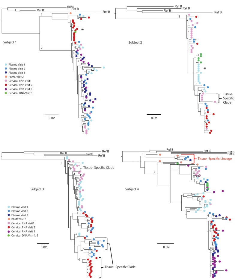
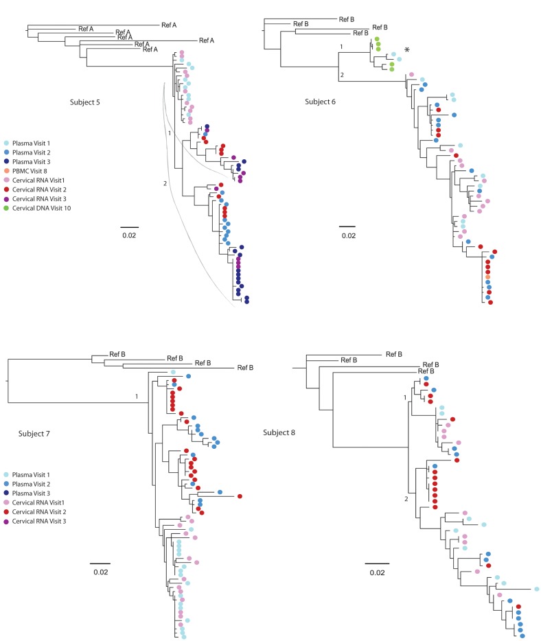
Methods: Eight women with ongoing HIV replication were studied during a period of 1.5 to 4.5 years. Multiple viral sequences were derived by single-genome amplification of the HIV C2-V5 region of env from genital secretions and blood plasma. Maximum likelihood phylogenies were evaluated for compartmentalization using 4 statistical tests.

Results: In cross-sectional analyses compartmentalization of genital from blood viruses was detected in three of eight women by all tests; this was associated with tissue specific clades containing multiple monotypic sequences. In longitudinal analysis, the tissues-specific clades did not persist to form viral lineages. Rather, across women, HIV lineages were comprised of both genital tract and blood sequences.

Conclusions: The observation of genital-specific HIV clades only in cross-sectional analysis and an absence of genital-specific lineages in longitudinal analyses suggest a dynamic interchange of HIV variants between the female genital tract and blood.


Figures

Figure 1.
Figure 1.
Maximum likelihood phylogenetic analyses of human immunodeficiency virus (HIV) RNA env C2-V5 from longitudinal specimens collected from the genital tract and blood of female subjects. HIV sequences were derived by single-genome amplification of complementary DNA derived from plasma (visit 1, light blue color; visit 2, darker blue; visit 3, darkest blue) and uterine cervical secretions (visit 1, light pink color; visit, 2 red; visit 3, fuschia). Sequences derived from CVL are indicated by light pink with a black circle (subject 4) but yielded significantly fewer sequences. Cell-associated sequences from PBMCs; (orange) or from cervical biopsies (green) are included from other visits in subjects 1, 2, 4, and 6 to demonstrate the lack of tissue-specific lineages but were not included in the analysis of compartmentalization. Tissue-specific lineages were limited to one subject: Subject 4, with a tissue-specific lineage in the peripheral blood. Tissue-specific clades are identified by brackets for subjects 1, 2, and 6; an * indicates tissue-specific HIV RNA clades before the addition of cell-associated sequences. The sequence predicted to encode X4 tropic virus is indicated by “X4.” The scale bar (horizontal line) indicates the branch length corresponding to 2 substitutions per 100 base pairs. Tissue-specific HIV env clades were composed of monotypic or of low-diversity sequences and were observed in all 8 subjects but were more pronounced in subjects 1–3, who had significant compartmentalization. The addition of cell-associated sequences to subjects 1, 2, and 6 demonstrated these clades were not restricted to 1 tissue and did not persist over time to form a “lineage” [29]. Genetically divergent sequences (>8% genetic distance) were noted in 3 women (subjects 1, 4, and 6) and were considered to possibly be due to intravaginal semen containing HIV or superinfection or significant evolution over time. Given the duration of these women's HIV infection (diagnosed 8, 17, and 12 years prior) and that HIV DNA from cervical biopsies or PBMCs from other study visits grouped in these clades, the latter 2 seem more likely.
Figure 1.
Figure 1.
Maximum likelihood phylogenetic analyses of human immunodeficiency virus (HIV) RNA env C2-V5 from longitudinal specimens collected from the genital tract and blood of female subjects. HIV sequences were derived by single-genome amplification of complementary DNA derived from plasma (visit 1, light blue color; visit 2, darker blue; visit 3, darkest blue) and uterine cervical secretions (visit 1, light pink color; visit, 2 red; visit 3, fuschia). Sequences derived from CVL are indicated by light pink with a black circle (subject 4) but yielded significantly fewer sequences. Cell-associated sequences from PBMCs; (orange) or from cervical biopsies (green) are included from other visits in subjects 1, 2, 4, and 6 to demonstrate the lack of tissue-specific lineages but were not included in the analysis of compartmentalization. Tissue-specific lineages were limited to one subject: Subject 4, with a tissue-specific lineage in the peripheral blood. Tissue-specific clades are identified by brackets for subjects 1, 2, and 6; an * indicates tissue-specific HIV RNA clades before the addition of cell-associated sequences. The sequence predicted to encode X4 tropic virus is indicated by “X4.” The scale bar (horizontal line) indicates the branch length corresponding to 2 substitutions per 100 base pairs. Tissue-specific HIV env clades were composed of monotypic or of low-diversity sequences and were observed in all 8 subjects but were more pronounced in subjects 1–3, who had significant compartmentalization. The addition of cell-associated sequences to subjects 1, 2, and 6 demonstrated these clades were not restricted to 1 tissue and did not persist over time to form a “lineage” [29]. Genetically divergent sequences (>8% genetic distance) were noted in 3 women (subjects 1, 4, and 6) and were considered to possibly be due to intravaginal semen containing HIV or superinfection or significant evolution over time. Given the duration of these women's HIV infection (diagnosed 8, 17, and 12 years prior) and that HIV DNA from cervical biopsies or PBMCs from other study visits grouped in these clades, the latter 2 seem more likely.

References

    1. Keele BF, Giorgi EE, Salazar-Gonzalez JF, et al. Identification and characterization of transmitted and early founder virus envelopes in primary HIV-1 infection. Proc Nat Acad Sci U S A. 2008;105:7552–7. - PMC - PubMed
    1. Gottlieb GS, Heath L, Nickle DC, et al. HIV-1 variation before seroconversion in men who have sex with men: analysis of acute/early HIV infection in the multicenter AIDS cohort study. J Infect Dis. 2008;197:1011–5. - PMC - PubMed
    1. Salazar-Gonzalez JF, Salazar MG, Keele BF, et al. Genetic identity, biological phenotype, and evolutionary pathways of transmitted/founder viruses in acute and early HIV-1 infection. J Exp Med. 2009;206:1273–89. - PMC - PubMed
    1. Kearney M, Maldarelli F, Shao W, et al. Human immunodeficiency virus type 1 population genetics and adaptation in newly infected individuals. J Virol. 2009;83:2715–27. - PMC - PubMed
    1. Fischer W, Ganusov VV, Giorgi EE, et al. Transmission of single HIV-1 genomes and dynamics of early immune escape revealed by ultra-deep sequencing. PLoS One. 5:e12303. - PMC - PubMed

Publication types

MeSH terms

Substances