. 2013 Mar;195(6):1294-304.
doi: 10.1128/JB.01997-12. Epub 2013 Jan 11.

Adaptive evolution of the Streptococcus pyogenes regulatory aldolase LacD.1

Affiliations

Adaptive evolution of the Streptococcus pyogenes regulatory aldolase LacD.1

Zachary Cusumano et al. J Bacteriol. 2013 Mar.

Abstract

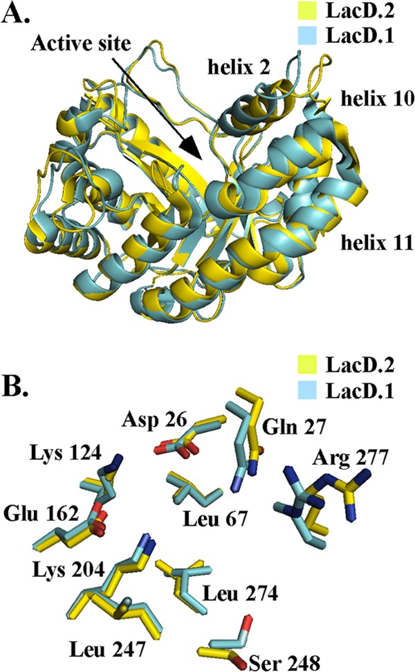
In the human-pathogenic bacterium Streptococcus pyogenes, the tagatose bisphosphate aldolase LacD.1 likely originated through a gene duplication event and was adapted to a role as a metabolic sensor for regulation of virulence gene transcription. Although LacD.1 retains enzymatic activity, its ancestral metabolic function resides in the LacD.2 aldolase, which is required for the catabolism of galactose. In this study, we compared these paralogous proteins to identify characteristics correlated with divergence and novel function. Surprisingly, despite the fact that these proteins have identical active sites and 82% similarity in amino acid sequence, LacD.1 was less efficient at cleaving both fructose and tagatose bisphosphates. Analysis of kinetic properties revealed that LacD.1's adaptation was associated with a decrease in k(cat) and an increase in K(m). Construction and analysis of enzyme chimeras indicated that non-active-site residues previously associated with the variable activities of human aldolase isoenzymes modulated LacD.1's affinity for substrate. Mutant LacD.1 proteins engineered to have LacD.2-like levels of enzymatic efficiency lost the ability to function as regulators, suggesting that an alteration in efficiency was required for adaptation. In competition under growth conditions that mimic a deep-tissue environment, LacD.1 conferred a significant gain in fitness that was associated with its regulatory activity. Taken together, these data suggest that LacD.1's adaptation represents a form of neofunctionalization in which duplication facilitated the gain of regulatory function important for growth in tissue and pathogenesis.


Figures

Fig 1
Fig 1
The crystal structures of LacD.1 (3MYO) and LacD.2 (3MHF) were compared. (A) Superposition of LacD.1's active site (cyan) and LacD.2's active site (yellow); active-site residues from LacD.1—D26, Q27, L67, K124, E162, K204, L247, S248, L274, and R277—are labeled. (B) Superposition of LacD.1 and LacD.2 crystal structures with thermal B-factors illustrated. The higher B-factors are associated with an increase in thickness.
Fig 2
Fig 2
The influence of pH on the enzyme efficiencies of both LacD.1 and LacD.2 was determined by measuring the Michaelis-Menten constants for cleavage of FBP at several different pHs in the range of 6.0 to 8.0. See Materials and Methods for details.
Fig 3
Fig 3
Gel filtration chromatography of LacD.1 and LacD.2. Purified LacD.1 and LacD.2 were subjected to gel filtration chromatography over Superdex 200 10/200 GL. Based on the elution of the standards (thyroglobin [669 kDa] [A], aldolase [158 kDa] [B], conalbumin [75 kDa] [C], ovalbumin [43 kDa] [D], and RNase A [13.7 kDa] [E]), the molecular masses of LacD.1 and LacD.2 were calculated to be 67 ± 3 and 71 ± 3 kDa, respectively.
Fig 4
Fig 4
Influence of enzyme concentration on enzyme activity. The FBP aldolase activities of LacD.1 and LacD.2 were measured at the enzyme concentrations 0.1, 0.325, 0.625, 1.25, 2.5, and 5 mM. Plotting of activity versus enzyme concentration revealed a linear relationship, with an R2 of 0.99 for both LacD.1 and LacD.2. See Materials and Methods for details.
Fig 5
Fig 5
Identification of polymorphic amino acid residues in LacD.1. (A) The polymorphic residues of LacD.1 (cyan) are mapped on the crystal structure of LacD.2. Catalytic residues identified above are highlighted in red. The locations of the loop 1 and turn 2 regions are labeled with arrows. (B) Identification of LacD.1 regions swapped with LacD.2; regions swapped are highlighted. LacD.12A includes red, green, and purple residues; LacD.12B includes green and purple residues; and LacD.12C includes purple residues.
Fig 6
Fig 6
Construction of LacD.1 chimeras. The amino acid sequence of LacD.2 is illustrated at the top, with black boxes representing the polymorphic residues between LacD.2 and LacD.1. The catalytic residues from LacD.1 are labeled. The LacD.1 sequence is indicated by the white box, and for each chimera, the black and white boxes represent the sequences derived from LacD.2 and LacD.1, respectively. The loop 1 and turn 2 sequences are highlighted below the boxes of selected chimeras, with the specific mutations introduced into the chimera underlined as represented by the sequence shown on the lower line. The name of each chimera is shown at the right.
Fig 7
Fig 7
Regulation of speB by LacD chimeras. (A) Spotting of overnight cultures on protease indicator media. Protease activity is apparent as a zone of clearing around the colony. The medium was unmodified (Unmod.) or buffered to a pH of 7.5 with the addition of 100 mM HEPES (pH 7.5). The HSC5 and LacD.1 strains contain empty vectors, while the other strains in a LacD.1 background contain either wild-type (WT) LacD.1, LacD.2, or the indicated chimeras described in Fig. 6. (B) Secreted-protease assay from supernatant fluid of the indicated strains described in Fig. 6. Buffered media were prepared as described in Materials and Methods. Activity is reported as a percentage of that of LacD.1. Data represent the means of at least three independent experiments, with samples analyzed in triplicate. An asterisk denotes a significant difference from LacD.1 pLacD.1/LacD.1 complemented with LacD chimeric enzymes in the same medium (P < 0.05).
Fig 8
Fig 8
Identification of regulatory regions of LacD.1. (A) Several additional LacD.1 and LacD.2 chimeras are shown and are represented as described for Fig. 6. (B) Protease activity of the indicated strains determined by a spotting assay on protease indicator media. The clear zone around the bacterial growth indicates production of proteolytic activity. (C) Quantitative determination of proteolytic activity in cell-free supernatant fluid of the indicated strains. Data represent the means of at least three independent experiments, with samples analyzed in triplicate.
Fig 9
Fig 9
The regulatory activity of LacD.1 confers increased fitness. Competition during growth in vitro was determined in coculture with a competitor strain (ΔLacD.1 complemented with pLacD.1−kan) and a test strain (ΔLacD.1 complemented with one of the various plasmids indicated). Cultures were back-diluted into fresh medium following 24 h of growth, and CFU were enumerated by plating on media with and without kanamycin for calculation of the competitive index. Data are the means of at least two independent experiments.

Similar articles

Cited by

References

    1. Kim JW, Dang CV. 2005. Multifaceted roles of glycolytic enzymes. Trends Biochem. Sci. 30:142–150 - PubMed
    1. Raffaelli N, Lorenzi T, Mariani PL, Emanuelli M, Amici A, Ruggieri S, Magni G. 1999. The Escherichia coli NadR regulator is endowed with nicotinamide mononucleotide adenylyltransferase activity. J. Bacteriol. 181:5509–5511 - PMC - PubMed
    1. Lohr D, Venkov P, Zlatanova J. 1995. Transcriptional regulation in the yeast GAL gene family: a complex genetic network. FASEB J. 9:777–787 - PubMed
    1. Platt A, Ross HC, Hankin S, Reece RJ. 2000. The insertion of two amino acids into a transcriptional inducer converts it into a galactokinase. Proc. Natl. Acad. Sci. U. S. A. 97:3154–3159 - PMC - PubMed
    1. Hahn MW. 2009. Distinguishing among evolutionary models for the maintenance of gene duplicates. J. Hered. 100:605–617 - PubMed

Publication types

MeSH terms

LinkOut - more resources