Review
. 2013 Feb 22;288(8):5443-50.
doi: 10.1074/jbc.R112.438382. Epub 2013 Jan 11.

Structure and dynamic regulation of Abl kinases

Affiliations
Review

Structure and dynamic regulation of Abl kinases

Shoghag Panjarian et al. J Biol Chem. .

Abstract

The c-abl proto-oncogene encodes a unique protein-tyrosine kinase (Abl) distinct from c-Src, c-Fes, and other cytoplasmic tyrosine kinases. In normal cells, Abl plays prominent roles in cellular responses to genotoxic stress as well as in the regulation of the actin cytoskeleton. Abl is also well known in the context of Bcr-Abl, the oncogenic fusion protein characteristic of chronic myelogenous leukemia. Selective inhibitors of Bcr-Abl, of which imatinib is the prototype, have had a tremendous impact on clinical outcomes in chronic myelogenous leukemia and revolutionized the field of targeted cancer therapy. In this minireview, we focus on the structural organization and dynamics of Abl kinases and how these features influence inhibitor sensitivity.


Figures

FIGURE 1.
FIGURE 1.
Domain organization of Abl kinases and crystal structure of the down-regulated c-Abl core. Upper, the c-Abl-1b protein kinase consists of a myristoylated (Myr) N-cap region, followed by SH3 and SH2 domains, the SH2/kinase linker, the tyrosine kinase domain, and a long last exon region with a C-terminal actin-binding domain (ABD). In Bcr-Abl, N-terminal fusion to Bcr sequences removes most of the N-cap, leaving the remainder of Abl intact. The Bcr-derived portion includes an N-terminal coiled-coil (CC) oligomerization domain as well as Dbl and pleckstrin homology domains (DH/PH). Lower, the x-ray crystal structure of the c-Abl-1b core protein in the down-regulated state (PDB code 2FO0) is rendered as a ribbon (lower left) and with the surface added (right) to emphasize the tight molecular packing of the regulatory regions (N-cap/SH3/SH2/linker) against the kinase domain. The unstructured portion of the myristoylated N-cap that engages the C-lobe of the kinase domain is shown as a dotted line. Domains in the structure are color-coded and correspond to the schematic shown at the top. In the kinase domain, the positions of helix αC and the activation loop (Act. Loop) are rendered in cyan and magenta, respectively. The side chain of the activation loop autophosphorylation site (Tyr412) is also shown.
FIGURE 2.
FIGURE 2.
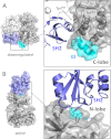
Reorientation of the SH2 domain as a function of Abl kinase activation. A, left, the position of the SH2 domain (blue) in the down-regulated structure of the c-Abl core is shown (PDB code 2FO0). Right, the interface of the SH2 domain with the back of the kinase domain is highlighted. The distal end of helix αI of the kinase domain C-lobe (cyan) is rotated away from the SH2 domain. Tyrosine residues from the SH2 (Tyr158) and kinase (Tyr361) domains form a pi-stacking interaction that contributes to the stabilization of the down-regulated conformation. B, the SH2 domain interacts with the N-lobe of the kinase domain to stabilize an active kinase domain conformation (PDB code 1OPL). Left, overview of this active top-hat conformation; right, close-up view of the SH2/N-lobe interface. Ile164 (cyan) from the SH2 domain plays a critical role in this interaction.
FIGURE 3.
FIGURE 3.
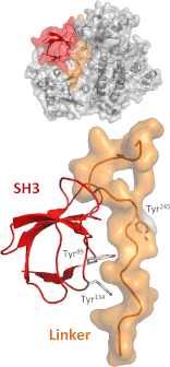
The Abl SH3 domain interacts with the SH2/kinase linker in down-regulated Abl. The positions of the SH3 domain (red) and the linker (orange) within the down-regulated c-Abl core structure (PDB code 2FO0) are shown (upper) and are also enlarged (lower). Note that the linker adopts a PPII helix that engages the SH3 domain. The side chains of three regulatory tyrosine phosphorylation sites are also shown. Two are located on the binding surface of the SH3 domain that faces the linker (Tyr89 and Tyr134), whereas the third is on the linker (Tyr245) and faces the N-lobe of the kinase domain.
FIGURE 4.
FIGURE 4.
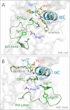
Structural features of the Abl kinase domain important for activity and inhibitor binding. A, close-up view of the Abl kinase domain bound to imatinib (PDB code 1IEP), which stabilizes a down-regulated conformation of the active site. Key structural elements of this off-state include the inward rotation of N-lobe helix αC (cyan), which positions Glu286 for ion pairing with Lys271. The DFG motif (Asp381, Phe382, and Gly383), which is located at the N-terminal end of the activation loop (Act Loop; green), is rotated away from the active site to accommodate imatinib binding (DFGout). The activation loop tyrosine (numbered as Tyr393 in this structure) points into the active site and makes a hydrogen bond with the catalytic aspartate (Asp363; purple). Also shown is the side chain of the gatekeeper residue (Thr315; orange), which forms a critical hydrogen bond with imatinib. B, close-up view of the Abl catalytic site in an active conformation with dasatinib bound (PDB code 2GQG; with ligand carbon atoms in yellow). Note that helix αC and the Glu286 ion pair with Lys271 are positioned as per the inactive state in A, but the activation loop is completely reoriented. The phosphorylated activation loop tyrosine (phospho-Tyr393) is now paired with a nearby arginine residue (Arg386), releasing the catalytic aspartate and stabilizing the active conformation. The DFG motif is now flipped inward (DFGin); this conformation is not compatible with imatinib binding due to steric clash. The gatekeeper threonine also makes a hydrogen bond with dasatinib.

Similar articles

Cited by

References

    1. Colicelli J. (2010) ABL tyrosine kinases: evolution of function, regulation, and specificity. Sci. Signal. 3, re6. - PMC - PubMed
    1. Hantschel O., Superti-Furga G. (2004) Regulation of the c-Abl and Bcr-Abl tyrosine kinases. Nat. Rev. Mol. Cell Biol. 5, 33–44 - PubMed
    1. Pendergast A. M. (2002) The Abl family kinases: mechanisms of regulation and signaling. Adv. Cancer Res. 85, 51–100 - PubMed
    1. Van Etten R. A. (1999) Cycling, stressed-out and nervous: cellular functions of c-Abl. Trends Cell Biol. 9, 179–186 - PubMed
    1. Pluk H., Dorey K., Superti-Furga G. (2002) Autoinhibition of c-Abl. Cell 108, 247–259 - PubMed

Publication types

MeSH terms