. 2013 Mar 1;288(9):6190-201.
doi: 10.1074/jbc.M112.426197. Epub 2013 Jan 11.

Deletion of vacuolar proton-translocating ATPase V(o)a isoforms clarifies the role of vacuolar pH as a determinant of virulence-associated traits in Candida albicans

Affiliations

Deletion of vacuolar proton-translocating ATPase V(o)a isoforms clarifies the role of vacuolar pH as a determinant of virulence-associated traits in Candida albicans

Summer M Raines et al. J Biol Chem. .

Abstract

Vacuolar proton-translocating ATPase (V-ATPase) is a central regulator of cellular pH homeostasis, and inactivation of all V-ATPase function has been shown to prevent infectivity in Candida albicans. V-ATPase subunit a of the Vo domain (Voa) is present as two fungal isoforms: Stv1p (Golgi) and Vph1p (vacuole). To delineate the individual contribution of Stv1p and Vph1p to C. albicans physiology, we created stv1Δ/Δ and vph1Δ/Δ mutants and compared them to the corresponding reintegrant strains (stv1Δ/ΔR and vph1Δ/ΔR). V-ATPase activity, vacuolar physiology, and in vitro virulence-related phenotypes were unaffected in the stv1Δ/Δ mutant. The vph1Δ/Δ mutant exhibited defective V1Vo assembly and a 90% reduction in concanamycin A-sensitive ATPase activity and proton transport in purified vacuolar membranes, suggesting that the Vph1p isoform is essential for vacuolar V-ATPase activity in C. albicans. The vph1Δ/Δ cells also had abnormal endocytosis and vacuolar morphology and an alkalinized vacuolar lumen (pHvph1Δ/Δ = 6.8 versus pHvph1Δ/ΔR = 5.8) in both yeast cells and hyphae. Secreted protease and lipase activities were significantly reduced, and M199-induced filamentation was impaired in the vph1Δ/Δ mutant. However, the vph1Δ/Δ cells remained competent for filamentation induced by Spider media and YPD, 10% FCS, and biofilm formation and macrophage killing were unaffected in vitro. These studies suggest that different virulence mechanisms differentially rely on acidified vacuoles and that the loss of both vacuolar (Vph1p) and non-vacuolar (Stv1p) V-ATPase activity is necessary to affect in vitro virulence-related phenotypes. As a determinant of C. albicans pathogenesis, vacuolar pH alone may prove less critical than originally assumed.


Figures

FIGURE 1.
FIGURE 1.
PCR verification of stv1Δ/Δ and vph1Δ/Δ strain construction in C. albicans. Homologous recombination was used to replace STV1 or VPH1 with URA3 (first allele) and ARG4 (second allele) to create the homozygous null C. albicans strains (Δ/Δ). A wild-type copy of the targeted gene was introduced on the pGEM-HIS1 plasmid to create the isogenic, complemented reintegrant strains (R). All strains were also compared with the DAY185 prototrophic control strain. DET primers spanned the targeted region of the disruption cassette, resulting in various sized bands depending on the allele present. The presence/absence of the target gene was confirmed with INT primers specific for an internal region of the open reading frame, and the presence of HIS1 was confirmed with HIS primers. The sequences of primers used are shown in Table 1.
FIGURE 2.
FIGURE 2.
The presence of a single Voa isoform is sufficient to maintain C. albicans growth. Growth is normal in both stv1Δ/Δ and vph1Δ/Δ mutant C. albicans strains across a range of pH. 5-Fold serial dilutions of overnight cultures were stamped onto pH-buffered complete synthetic media plates and grown for 48–72 h at 30 °C.
FIGURE 3.
FIGURE 3.
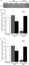
Vacuolar ATPase activity is drastically reduced in C. albicans vph1Δ/Δ mutants. A, VoV1 V-ATPase complexes are not properly assembled at the vacuolar membrane in vph1Δ/Δ mutants. Purified vacuolar vesicles were analyzed by Western blot using an anti-V1A antibody. A representative image (from three blots of three independent vacuolar preps) is shown. B, ATP hydrolysis is dramatically reduced in vph1Δ/Δ mutants. Concanamycin-A-sensitive ATP hydrolysis of purified vacuolar vesicles was measured spectrophotometrically using an enzyme assay coupled to NADH oxidation at 340 nm. The average DAY185-specific activity was 0.95 μmol of Pi/min/mg. C, proton transport is dramatically reduced in vph1Δ/Δ mutants. ATP-dependent proton transport of purified vacuolar vesicles was measured via fluorescence quenching of 1 μm 9-amino-6-chloro-2-methoxyacridine upon the addition of 0.5 mm ATP, 1 mm MgSO4. Proton transport was calculated as the change in fluorescence for the first 15 s after ATP addition. The average DAY185 slope was −1340.75 fluorescence units/15 s. Percentage activities are expressed relative to DAY185 and are shown as the average ± S.D. for n = 3–6 separate vacuolar purifications; percent reductions in activity are indicated. **, p < 0.01; ****, p < 0.0001 versus the reintegrant strain (R) as measured by a two-tailed unpaired Student's t test.
FIGURE 4.
FIGURE 4.
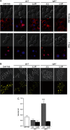
Vacuolar biogenesis is disrupted in C. albicans vph1Δ/Δ mutants. A, endocytosis to the vacuole and vacuolar morphology was impaired in vph1Δ/Δ mutants. Live C. albicans cells were stained with FM4-64 dye (red) and CMAC dye (blue) to mark the vacuolar membrane and the vacuolar lumen, respectively. Representative differential interference contrast and fluorescence images are shown. FM4-64 internalization to the vacuole was assessed after 15 min and after 1 h. B and C, the vacuolar lumen is alkalinized in vph1Δ/Δ mutants. Quinacrine accumulation (B) was used to qualitatively assess vacuolar acidification in live C. albicans cells; quinacrine fluorescence is quenched as pH increases. Representative differential interference contrast and fluorescent images are shown. BCECF accumulation (C) was used to quantitatively assess vacuolar pH in live C. albicans cells; BCECF fluorescence increases with pH. Cells were stained with 50 μm BCECF-AM for 30 min, and the average fluorescence over 7 min was compared with a standard curve to generate absolute pH values. Vacuolar pH is expressed as the average ± S.D. for n = 3–6 replicates. ****, p < 0.0001 versus the reintegrant strain (R) as measured by a two-tailed unpaired Student's t test.
FIGURE 5.
FIGURE 5.
Secreted protease and lipase activity are reduced in C. albicans vph1Δ/Δ mutants. Extracellular aspartyl protease and lipase secretion/activity are reduced in vph1Δ/Δ mutants. For all assays, overnight cultures were spotted onto plates and grown at the specified time and temperature. A, proteases were assayed via BSA degradation plate assay. B, lipases were assayed on yeast nitrogen base plates containing 2.5% Tween 80. Representative images are shown for all assays. The sap1–3Δ/Δ (48) and sur7Δ/Δ (46) strains are included as negative controls for protease and lipase secretion, respectively. The amount of protease (C) and the amount of lipase secretion/activity (D) were quantified by measuring the diameter of the degradation halo surrounding the fungal colony, as halo size is proportional to the extracellular enzyme activity present. Halo diameters were normalized to the diameter of the colony itself; therefore, a ratio of 1 indicates a lack of detectable halo. Halo size is shown as the average ± S.E. for n = 8–16 replicates. *, p < 0.05; **, p < 0.01 versus the reintegrant strain (R) as measured by a two-tailed unpaired Student's t test.
FIGURE 6.
FIGURE 6.
In vitro virulence-related traits are differentially altered in C. albicans vph1Δ/Δ mutants. A, filamentation specifically in response to M199 media and agar embedding is reduced in vph1Δ/Δ mutants. Filamentation was assayed on various hyphae-inducing solid medias. For the embedded agar assays, cells were mixed with YPD agar before plates were poured. For all other assays, overnight cultures were spotted onto plates and grown at the times and temperatures indicated. Representative images are shown for all assays. B, biofilm formation is normal in vph1Δ/Δ mutants. Biofilm formation was calculated as the A490 measured using the XTT (2′,3′-bis-(2-methoxy-4-nitro-5-sulfophenyl)-5-(phenylaminocarbonyl)-2H-tetrazolium hydroxide) reduction method. C, macrophage killing is normal in vph1Δ/Δ mutants. Macrophages (J774A.1) were co-incubated with C. albicans strains for 24 h. Macrophage killing is inversely proportional to the number of live macrophages (as assessed by positive staining for calcein AM) counted from four microscopic fields per replicate. Biofilm formation is shown as the average ± S.D. for n = 8 replicates. Macrophage survival is shown as the average ± S.E. of three experiments, with n = 10–12 replicates per experiment.
FIGURE 7.
FIGURE 7.
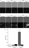
Induction of hyphal formation does not rescue vacuolar pH in vph1Δ/Δ C. albicans. A–C, C. albicans filamentation occurs despite elevated vacuolar pH. Hyphae formation was induced over 24 h in liquid YPD +10% FCS. Quinacrine accumulation into hyphae was used to qualitatively assess vacuolar pH during filamentation as described in the legend to Fig. 4B. Representative differential interference contrast and fluorescent images are shown at 4 h (A) and 24 h (B) post-hyphae induction; results were similar at 1 and 2 h.3 BCECF accumulation into hyphae was used to quantitatively assess vacuolar pH during filamentation as described in the legend to Fig. 4C. Vacuolar pH was calculated at 24 h post-hyphae induction (C) and is expressed as the average ± S.D. for n = 3–6 separate readings. ****, p < 0.0001 versus the reintegrant strain (R) as measured by a two-tailed unpaired Student's t test.

Similar articles

Cited by

References

    1. Toei M., Saum R., Forgac M. (2010) Regulation and isoform function of the V-ATPases. Biochemistry 49, 4715–4723 - PMC - PubMed
    1. Kane P. M. (2006) The where, when, and how of organelle acidification by the yeast vacuolar H+-ATPase. Microbiol. Mol. Biol. Rev. 70, 177–191 - PMC - PubMed
    1. Manolson M. F., Proteau D., Jones E. W. (1992) Evidence for a conserved 95–120-kDa subunit associated with and essential for activity of V-ATPases. J. Exp. Biol. 172, 105–112 - PubMed
    1. Manolson M. F., Proteau D., Preston R. A., Stenbit A., Roberts B. T., Hoyt M. A., Preuss D., Mulholland J., Botstein D., Jones E. W. (1992) The VPH1 gene encodes a 95-kDa integral membrane polypeptide required for in vivo assembly and activity of the yeast vacuolar H+-ATPase. J. Biol. Chem. 267, 14294–14303 - PubMed
    1. Kawasaki-Nishi S., Nishi T., Forgac M. (2001) Arg-735 of the 100-kDa subunit a of the yeast V-ATPase is essential for proton translocation. Proc. Natl. Acad. Sci. U.S.A. 98, 12397–12402 - PMC - PubMed

Publication types

LinkOut - more resources