. 2013 Mar 15;304(6):H828-39.
doi: 10.1152/ajpheart.00752.2012. Epub 2013 Jan 11.

Oxidative activation of Ca(2+)/calmodulin-activated kinase II mediates ER stress-induced cardiac dysfunction and apoptosis

Affiliations

Oxidative activation of Ca(2+)/calmodulin-activated kinase II mediates ER stress-induced cardiac dysfunction and apoptosis

Nathan D Roe et al. Am J Physiol Heart Circ Physiol. .

Abstract

Endoplasmic reticulum (ER) stress elicits oxidative stress and intracellular Ca(2+) derangement via activation of Ca(2+)/calmodulin-dependent protein kinase II (CaMKII). This study was designed to examine the role of CaMKII in ER stress-induced cardiac dysfunction and apoptosis as well as the effect of antioxidant catalase. Wild-type FVB and transgenic mice with cardiac-specific overexpression of catalase were challenged with the ER stress inducer tunicamycin (3 mg/kg ip for 48 h). Presence of ER stress was verified using the ER stress protein markers immunoglobulin binding protein (BiP) and C/EBP homologous protein (CHOP), the effect of which was unaffected by catalase overexpression. Echocardiographic assessment revealed that tunicamycin elicited cardiac remodeling (enlarged end-systolic diameter without affecting diastolic and ventricular wall thickness), depressed fractional shortening, ejection fraction, and cardiomyocyte contractile capacity, intracellular Ca(2+) mishandling, accumulation of reactive oxygen species (superoxide production and NADPH oxidase p47phox level), CaMKII oxidation, and apoptosis (evidenced by Bax, Bcl-2/Bax ratio, and TUNEL staining), the effects of which were obliterated by catalase. Interestingly, tunicamycin-induced cardiomyocyte mechanical anomalies and cell death were ablated by the CaMKII inhibitor KN93, in a manner reminiscent of catalase. These data favored a permissive role of oxidative stress and CaMKII activation in ER stress-induced cardiac dysfunction and cell death. Our data further revealed the therapeutic potential of antioxidant or CaMKII inhibition in cardiac pathological conditions associated with ER stress. This research shows for the first time that contractile dysfunction caused by ER stress is a result of the oxidative activation of the CaMKII pathway.


Figures
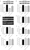
Fig. 1.
Fig. 1.
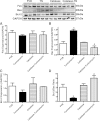
Effect of cardiac-specific overexpression of catalase on tunicamycin (3 mg/kg ip for 48 h)-induced endoplasmic reticulum (ER) stress and changes in echocardiographic indexes. A: BiP expression. B: CHOP expression. Insets in A and B: representative gel blots depicting expression of BiP, CHOP, and α-tubulin (loading control). C: representative echocardiographic images from FVB and catalase mice treated with or without tunicamycin. D: left ventricular (LV) wall thickness. E: LV end-diastolic diameter (LVEDD). F: LV end-systolic diameter (LVESD). G: fractional shortening. H: ejection fraction. Values are means ± SE; n = 5–7 mice/group. *P < 0.05 vs. FVB. #P < 0.05 vs. tunicamycin group.
Fig. 2.
Fig. 2.
Effect of cardiac-specific overexpression of catalase on tunicamycin (3 mg/kg ip for 48 h)-induced changes in cardiomyocyte contractile properties. A: representative cell shortening traces. B: peak shortening amplitude (normalized to cell length). C: maximum velocity of shortening (+dL/dt). D: maximum velocity of relengthening (−dL/dt). E: time to shortening (TPS). F: time to 90% relengthening (TR90). Values are means ± SE; n = 141–150 cells per group, *P < 0.05 vs. FVB. #P < 0.05 vs. tunicamycin group.
Fig. 3.
Fig. 3.
Effect of cardiac-specific overexpression of catalase on tunicamycin (TN, 3 mg/kg ip for 48 h)-induced changes in apoptotic proteins Bax, Bcl-2, and Fas. A: Fas expression. B: Bax expression. C: Bcl-2 expression. D: Bax-to-Bcl-2 ratio. Inset: gel blots of Fas, Bax, Bcl-2, and GAPDH (loading control). TN, tunicamycin. Values are means ± SE; n = 3 hearts per group. *P < 0.05 vs. FVB. #P < 0.05 vs. tunicamycin group.
Fig. 4.
Fig. 4.
TUNEL staining of myocardium from FVB and cardiac-specific catalase overexpression mice with or without tunicamycin (3 mg/kg ip for 48 h) challenge. A: representative images of TUNEL staining (left column) and DAPI nuclear labeling (right column). Arrows are pointing to apoptotic nuclei. B: quantitative analysis of TUNEL positive nuclei per 1,000 nuclei. Values are means ± SE; n = 3–4 hearts per group. *P < 0.05 vs. FVB. #P < 0.05 vs. tunicamycin group.
Fig. 5.
Fig. 5.
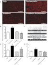
Effect of cardiac-specific overexpression of catalase on tunicamycin (3 mg/kg ip for 48 h)-induced changes in superoxide production, NADPH oxidase p47phox level, and oxidation of Ca2+/calmodulin-activated kinase II (CaMKII). A: representative images of DHE-stained heart sections. B: pooled data summarizing DHE fluorescence intensity. C: representative gel blots depicting p47phox, oxidized CaMKII, and GAPDH (loading control). D: p47phox expression. E: levels of oxidized CaMKII assessed using Western blot analysis. Values are means ± SE; n = 3–4 hearts per group. *P < 0.05 vs. FVB. #P < 0.05 vs. tunicamycin group.
Fig. 6.
Fig. 6.
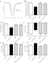
Effect of CaMKII inhibition on endoplasmic reticulum (ER) stress-induced cardiomyocyte contractile dysfunction. Freshly isolated cardiomyocytes from FVB control mice were incubated with tunicamycin (3 μg/ml) for 3 h in the presence or absence of the CaMKII inhibitor KN93 (0.5 μM) prior to assessment of cardiomyocyte mechanical function. A: representative cell shortening traces. B: peak shortening. C: +dL/dt. D: −dL/dt. E: TPS. F: TR90. Values are means ± SE; n = 116–118 cells per group. *P < 0.05 vs. control. #P < 0.05 vs. tunicamycin group.
Fig. 7.
Fig. 7.
Effect of catalase overexpression or CaMKII inhibition on ER stress-induced intracellular Ca2+ homeostasis. FVB and catalase mice were challenged with tunicamycin (3 mg/kg ip) prior to assessment of intracellular Ca2+ handling. A cohort of cardiomyocytes from FVB mice with or without tunicamycin treatment were incubated with the CaMKII inhibitor KN93 (0.5 μM, 30-min pretreatment) prior to intracellular Ca2+ fluorescence assessment. A: representative traces from FVB cardiomyocytes with or without tunicamycin treatment. B: resting fura-2 fluorescence intensity (FFI). C: electronically stimulated rise in FFI (ΔFFI). D: intracellular Ca2+ decay rate. Values are means ± SE; n = 55–61 cells per group. *P < 0.05 vs. FVB. #P < 0.05 vs. tunicamycin group.
Fig. 8.
Fig. 8.
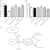
Effect of catalase overexpression or CaMKII inhibition on ER stress-induced apoptosis and change in mitochondrial membrane potential. FVB and catalase mice were challenged with tunicamycin (3 mg/kg ip) prior to biochemical assessment. A cohort of cardiomyocytes from FVB mice with or without tunicamycin treatment were incubated with the CaMKII inhibitor KN93 (0.5 μM, 30-min pretreatment) prior to biochemical assessment. A: cell viability (n = 8 isolations). B: mitochondrial membrane potential using JC-1 fluorescence (n = 15 fields). C: schematic diagram depicting proposed mechanism responsible for ER stress-induced cardiac dysfunction and apoptosis. ER stress triggers reactive oxygen species (ROS) production leading to oxidation of CaMKII, en route to cardiac dysfunction and apoptosis. Catalase prevents oxidation of CaMKII whereas KN93 inhibits CaMKII activation to alleviate cardiac dysfunction and apoptosis under ER stress. Values are means ± SE. *P < 0.05 vs. FVB. #P < 0.05 vs. tunicamycin.

Similar articles

Cited by

References

    1. Amanso AM, Debbas V, Laurindo FR. Proteasome inhibition represses unfolded protein response and Nox4, sensitizing vascular cells to endoplasmic reticulum stress-induced death. PLoS One 6: e14591, 2011 - PMC - PubMed
    1. Anderson ME, Brown JH, Bers DM. CaMKII in myocardial hypertrophy and heart failure. J Mol Cell Cardiol 51: 468–473, 2011 - PMC - PubMed
    1. Baines CP. The cardiac mitochondrion: nexus of stress. Annu Rev Physiol 72: 61–80, 2010 - PubMed
    1. Bers DM. CaMKII inhibition in heart failure makes jump to human. Circ Res 107: 1044–1046, 2010 - PubMed
    1. Bers DM, Grandi E. Calcium/calmodulin-dependent kinase II regulation of cardiac ion channels. J Cardiovasc Pharmacol 54: 180–187, 2009 - PMC - PubMed

Publication types

MeSH terms

LinkOut - more resources