. 2013 Mar 15;304(6):L394-400.
doi: 10.1152/ajplung.00091.2012. Epub 2013 Jan 11.

Increased expression of senescence markers in cystic fibrosis airways

Affiliations

Increased expression of senescence markers in cystic fibrosis airways

Bernard M Fischer et al. Am J Physiol Lung Cell Mol Physiol. .

Abstract

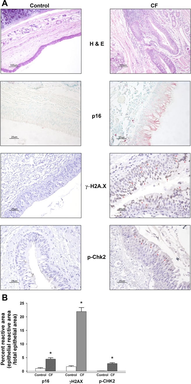
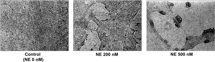
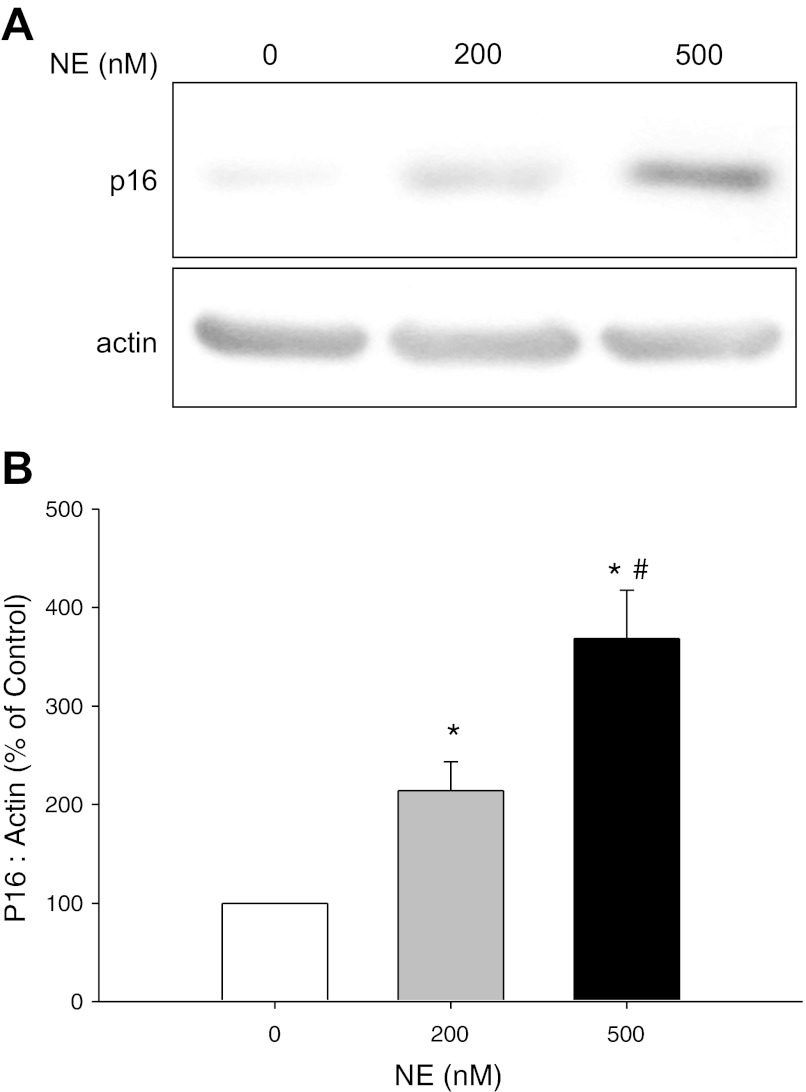
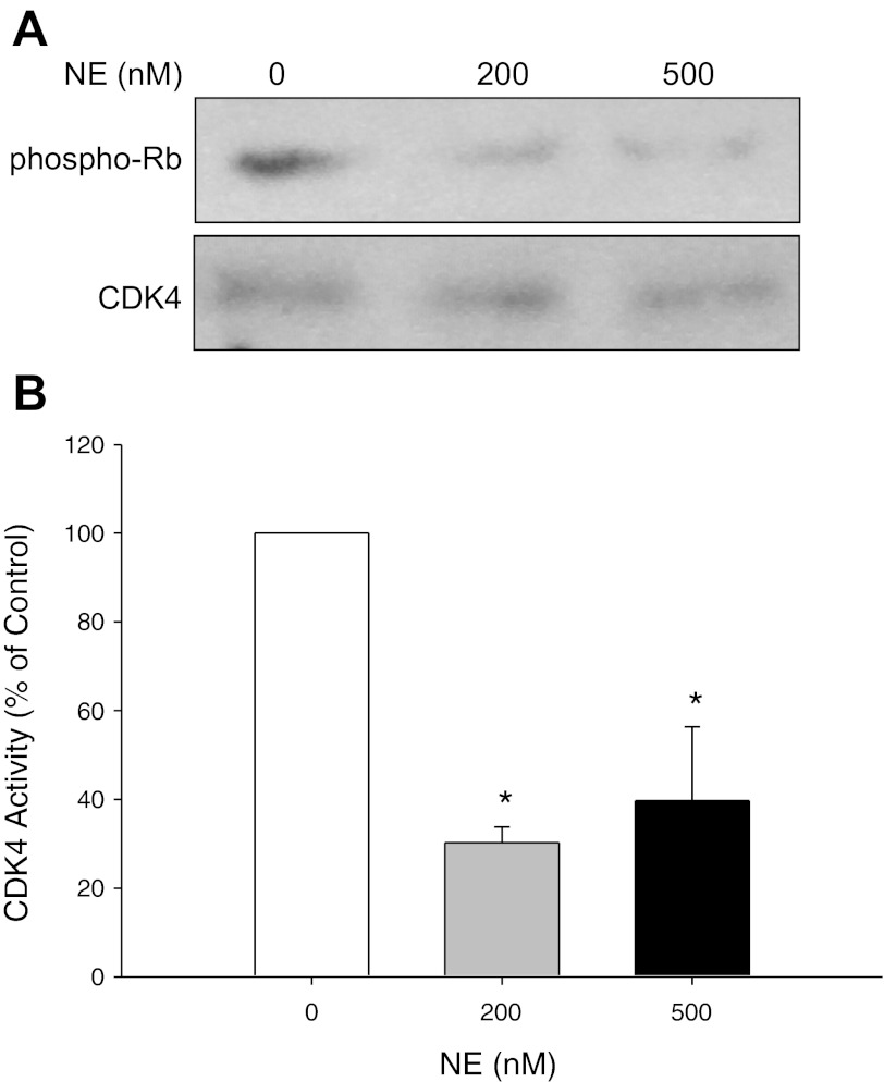
Cystic Fibrosis (CF) is a chronic lung disease characterized by chronic neutrophilic airway inflammation and increased levels of neutrophil elastase (NE) in the airways. We have previously reported that NE treatment triggers cell cycle arrest. Cell cycle arrest can lead to senescence, a complete loss of replicative capacity. Importantly, senescent cells can be proinflammatory and would perpetuate CF chronic inflammation. By immunohistochemistry, we evaluated whether airway sections from CF and control subjects expressed markers of senescence, including p16(INK4a) (p16), a cyclin-dependent kinase inhibitor, phospho-Histone H2A.X (γH2A.X), and phospho-checkpoint 2 kinase (phospho-Chk2), which are also DNA damage response markers. Compared with airway epithelium from control subjects, CF airway epithelium had increased levels of expression of all three senescence markers. We hypothesized that the high load of NE in the CF airway triggers epithelial senescence by upregulating expression of p16, which inhibits cyclin-dependent kinase 4 (CDK4). Normal human bronchial epithelial (NHBE) cells, cultured in air-liquid interface were treated with NE (0, 200, and 500 nM) to induce visible injury. Total cell lysates were collected and evaluated by Western analysis for p16 protein expression and CDK4 kinase activity. NE significantly increased p16 expression and decreased CDK4 kinase activity in NHBE cells. These results support the concept that NE triggers expression of senescence markers in CF airway epithelial cells.


Figures

Fig. 1.
Fig. 1.
Immunohistochemistry for p16INK4, phospho-Histone H2A.X (γ-H2AX), and phospho-Chk2 (p-Chk2) in control and cystic fibrosis (CF) airway tissue sections. A: representative hematoxylin and eosin (H&E) images (×10 magnification, 100 μm bar) from control (n = 12) and CF (n = 9). Note the increased cellularity of the CF airway epithelium and submucosal area indicative of inflammatory cell infiltration. Control (n = 6) and CF (n = 6) airway tissue sections (5 μm) were immunostained for senescence biomarker cyclin-dependent kinase (CDK) inhibitor, p16INK4 (p16) (alkaline phosphatase detection, pink color with methyl green counterstain), phosphorylated histone 2AX (γ-H2AX; control n = 5, CF n = 7) [diaminobenzidine (DAB), brown color with hematoxylin counterstain], and phospho-Chk2 (p-Chk2; n = 7 for each control and CF) (DAB, brown color with hematoxylin counterstain). Red arrow heads in γ-H2AX and p-Chk2 CF images indicate examples of positively staining nuclei in the epithelium. All senescence marker images were taken at ×40 magnification (25 μm bar) as noted in materials and methods. According to antibody supplier (BD Biosciences), the p16 monoclonal antibody will recognize p16 expression in both the cytoplasm and the nucleus. B: graphic summary of the unbiased quantitative histological analysis for each of the immunostained senescence/DNA damage markers (means ± SE; n = 5–17 images per airway from each control and CF subject). Results are expressed as percentage reactive (stained) area = ratio of epithelial reactive (stained) area to total epithelial area. *CF significantly increased compared with corresponding control subjects stained airways, P < 0.05.
Fig. 2.
Fig. 2.
Absolute telomere length (aTL) of CF and control airway epithelial cell genomic DNA. DNA was extracted from both control (CTRL) and CF passage 1 airway epithelial cells and analyzed by qPCR for telomere DNA and normalized to single copy gene (SCG) 36B4 (n = 18 per group). On the basis of a standard curve for each telomere and 36B4 DNA, the total telomere length in kb per human diploid genome was calculated. The result was then divided by 92 (total number of telomeres on 23 pairs of human chromosomes) to yield the length per telomere or aTL. A and B: standard curves using oligomer standards for telomere DNA (TEL), 84 bp oligomer, and 36B4 SCG, 75 bp oligomer, were used to calculate the telomere length for each sample. Graphs are plotted with qPCR CT (cycle threshold) as the y-axis. The x-axis is the Log of kb of telomere DNA (TEL) represented by each standard (A) or the Log of 36B4 SCG diploid genome copies represented by each standard (B). These graphs demonstrate the consistent repeatability of the standard curves for this method. C: box-and-whisker plot of CTRL and CF aTL. The line in the middle of each box represents the median. The “whiskers” represent the spread of the data with no outliers noted. D: scatter plot of CTRL and CF aTL. The line represents the median in each group. The three samples circled are those patients with CF whose aTL was below all the control subjects.
Fig. 3.
Fig. 3.
Neutrophil elastase (NE)-induced injury of normal human bronchial epithelial (NHBE) cells grown in air-liquid interface (ALI) cultures. NHBE were treated with neutrophil elastase (NE; 0, 200, or 500 nM) to induce visible injury. In the absence of NE (0 nM), NHBE form a confluent, differentiated cell monolayer. NE 200 nM exhibits small holes of injury in the cell monolayer. In cells exposed to NE 500 nM, the monolayer is completely disrupted.
Fig. 4.
Fig. 4.
Western analysis of p16 INK4 protein expression in NHBE cells. NHBE cells grown in ALI culture were treated with control vehicle or NE (0, 200, or 500 nM) for 2–5 h until visible injury occurred. At the end of the treatment period, cell lysates were collected and separated (∼80 μg total protein) on a 4–20% SDS-PAGE. After transfer to nitrocellulose, membrane was probed with a monoclonal antibody for p16INK4 (p16). A monoclonal antibody for β-actin was used to demonstrate equivalent protein loading. A: representative autoradiographs. B: graphic summary of densitometric analysis of autoradiographs from 6 experiments (means ± SE; n = 7–8). Results were expressed as a ratio of p16 protein expression to β-actin protein expression and then expressed as a percentage of the corresponding control. *NE 200 and 500 nM significantly greater than control (NE 0 nM), P < 0.05. #NE 500 nM significantly greater than NE 200 nM, P < 0.05.
Fig. 5.
Fig. 5.
Western analysis of CDK4 activity in NHBE cells. NHBE cells grown in ALI culture were treated with control vehicle or NE (0, 200, or 500 nM) for 2–5 h until visible injury occurred. At the end of the treatment period, cell lysates were immunoprecipitated (IP) with a mouse monoclonal anti-human antibody to CDK4. Protein complexes were collected with Dynabeads Protein G and then incubated with truncated His-Tagged Retinoblastoma protein (Rb) substrate (1 μg) in kinase reaction buffer. Samples were then separated on a 4–20% SDS-PAGE, transferred to nitrocellulose, and the membrane probed with anti-phospho-pRB (Ser 795) antibody (1:1,000) to measure CDK4 activity phosphorylated at the serine 795 site. An anti-CDK4 polyclonal rabbit antibody (1:1,000) was used to measure total CDK4 protein levels to demonstrate that NE did not change total CDK4 levels and equivalent CDK4 protein was immunoprecipitated. A: representative autoradiographs. B: graphic summary of densitometric analysis of autoradiographs from 3 experiments (means ± SE; n = 4). *NE 200 and 500 nM significantly less than control (NE 0 nM), P < 0.05.
Fig. 6.
Fig. 6.
Heterogeneity of airway epithelial cell fates in response to NE. NE regulates several alternative airway epithelial cell fates: cell cycle arrest (15), apoptosis (16, 37), and senescence. In this report, we demonstrate that NE upregulated p16 and inhibited CDK4; this pathway induces epithelial senescence.

Similar articles

Cited by

References

    1. Aoshiba K, Yasuda K, Yahui S, Tamaoki J, Nagai A. Serine proteases increase oxidative stress in lung cells. Am J Physiol Lung Cell Mol Physiol 281: L556–L564, 2001 - PubMed
    1. Barash H, ERG , Edrei Y, Ella E, Israel A, Cohen I, Corchia N, Ben-Moshe T, Pappo O, Pikarsky E, Goldenberg D, Shiloh Y, Galun E, Abramovitch R. Accelerated carcinogenesis following liver regeneration is associated with chronic inflammation-induced double-strand DNA breaks. Proc Natl Acad Sci USA 107: 2207–2212, 2010 - PMC - PubMed
    1. Birrer P, McElvaney NG, Rudeberg A, Sommer CW, Liechti-Gallati S, Kraemer R, Hubbard R, Crystal RG. Protease-antiprotease imbalance in the lungs of children with cystic fibrosis. Am J Respir Crit Care Med 150: 207–213, 1994 - PubMed
    1. Brennan S, Sly PD, Gangell CL, Sturges N, Winfield K, Wikstrom M, Gard S, Upham JW. Alveolar macrophages and CC chemokines are increased in children with cystic fibrosis. Eur Respir J 34: 655–661, 2009 - PubMed
    1. Britigan BE, Edeker BL. Pseudomonas and neutrophil products modify transferrin and lactoferrin to create conditions that favor hydroxyl radical formation. J Clin Invest 88: 1092–1102, 1991 - PMC - PubMed

Publication types

MeSH terms