. 2013 Mar 15;304(6):L415-27.
doi: 10.1152/ajplung.00180.2012. Epub 2013 Jan 11.

Role of β-catenin-regulated CCN matricellular proteins in epithelial repair after inflammatory lung injury

Affiliations

Role of β-catenin-regulated CCN matricellular proteins in epithelial repair after inflammatory lung injury

Rachel L Zemans et al. Am J Physiol Lung Cell Mol Physiol. .

Abstract

Repair of the lung epithelium after injury is integral to the pathogenesis and outcomes of diverse inflammatory lung diseases. We previously reported that β-catenin signaling promotes epithelial repair after inflammatory injury, but the β-catenin target genes that mediate this effect are unknown. Herein, we examined which β-catenin transcriptional coactivators and target genes promote epithelial repair after inflammatory injury. Transmigration of human neutrophils across cultured monolayers of human lung epithelial cells resulted in a fall in transepithelial resistance and the formation of discrete areas of epithelial denudation ("microinjury"), which repaired via cell spreading by 96 h. In mice treated with intratracheal (i.t.) LPS or keratinocyte chemokine, neutrophil emigration was associated with increased permeability of the lung epithelium, as determined by increased bronchoalveolar lavage (BAL) fluid albumin concentration, which decreased over 3-6 days. Activation of β-catenin/p300-dependent gene expression using the compound ICG-001 accelerated epithelial repair in vitro and in murine models. Neutrophil transmigration induced epithelial expression of the β-catenin/p300 target genes Wnt-induced secreted protein (WISP) 1 and cysteine-rich (Cyr) 61, as determined by real-time PCR (qPCR) and immunostaining. Purified neutrophil elastase induced WISP1 upregulation in lung epithelial cells, as determined by qPCR. WISP1 expression increased in murine lungs after i.t. LPS, as determined by ELISA of the BAL fluid and qPCR of whole lung extracts. Finally, recombinant WISP1 and Cyr61 accelerated repair, and Cyr61-neutralizing antibodies delayed repair of the injured epithelium in vitro. We conclude that β-catenin/p300-dependent expression of WISP1 and Cyr61 is critical for epithelial repair and represents a potential therapeutic target to promote epithelial repair after inflammatory injury.


Figures

Fig. 1.
Fig. 1.
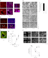
Inflammatory injury followed by repair of the lung epithelium in vitro. AH: neutrophils were induced to migrate across Calu-3 or Calu-3-green fluorescent protein (GFP) cells for 120 min by a gradient of N-Formyl-Met-Leu-Phe (fMLP) (1 μM). A: transepithelial resistance (TER) was measured. B and C: fluorescent images (×2.5) were acquired at the specified time points, and the total cross-sectional area of epithelial defects in 5 randomly selected high-powered fields (HPF) was measured. Arrowheads indicate epithelial wounds. D: at 30 h after migration, epithelial monolayers were cultured with 10 μM 5-bromo-2-deoxyuridine (BrdU) for 2 h, fixed, and stained with anti-BrdU antibodies. The dashed circle indicates an epithelial wound. E: at 30 h after migration, epithelial monolayers were fixed and stained with phalloidin and DAPI. Closed arrows indicate enhanced F-actin staining in the leading edge of cells migrating into the denuded area; open arrows indicate baseline F-actin staining in cells that do not appear to be migrating into the denuded area. The boxed region indicates the area magnified in the inset (right). F: at 30 h after migration, epithelial monolayers were fixed and stained with DAPI. Fixed monolayers were analyzed for internuclear distances. At 24 h after migration, epithelial monolayers were cultured with 1 μg/ml cytochalasin (G) or 1 μg/ml mitomycin (H). In G, area of denudation at 48 h is expressed as a percentage of the area of initial injury at 24 h, with the control group expressed as a percentage of the treatment group. Fluorescent images (×2.5) were acquired at the specified time points, and the total cross-sectional area of epithelial defects in 5 randomly selected HPF was measured. I: Calu-3 cells were seeded into 24-well plates, treated with 1 μg/ml mitomycin for 3 days, and trypsinized and counted. n ≥ 4. *P ≤ 0.05. Error bars represent SE.
Fig. 2.
Fig. 2.
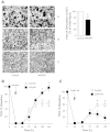
Inflammatory injury followed by repair of the lung epithelium in vivo. C57BL/6 mice were treated with LPS (A) 20 μg in 50 μl saline or 50 μl saline or keratinocyte chemokine (KC) (B) 1 μg in 50 μl saline + 0.1% human serum albumin (HSA) or 50 μl 0.1% HSA i.t. and euthanized at the indicated time points. Bronchoalveolar lavage (BAL) cell count and differential were performed, and albumin concentrations were measured by ELISA. Error bars represent SE.
Fig. 3.
Fig. 3.
β-Catenin/p300 signaling accelerates repair of the lung epithelium after inflammatory injury and mechanical wounding in vitro. Neutrophils were induced to migrate across Calu-3 (AC) or Calu-3-GFP (D and E) cells for 90–120 min. At 24 h after migration, the epithelial monolayer was fixed and stained for active β-catenin (A) or p300 (B) or active β-catenin and p300 (C). Arrows indicate nuclear β-catenin and/or p300. Boxed region indicates the area magnified in the inset (right). D and E: ICG-001 was added to the culture media at 24 h after migration, as described in materials and methods. At the indicated time points, TER (D) was measured, and fluorescent images (×2.5) (E) were acquired. The total cross-sectional area of epithelial defects in 5 randomly selected HPF was measured. Arrowheads indicate epithelial wounds. Area is normalized to control in each experiment. In these experiments, the media were changed at 24 h after migration to remove secreted growth factors, which likely prevented the TER from returning to baseline in the control samples. F: scratch wounds were made on confluent monolayers of Calu-3-GFP cells, followed immediately by the addition of ICG-001 to the culture media. The area of defect within a 5-mm2 region centered on the intersection of the scratches was measured. *P < 0.05; n ≥ 4. Error bars represent SE.
Fig. 4.
Fig. 4.
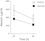
β-catenin/p300 signaling accelerates repair of the lung epithelium after inflammatory injury in murine models. C57BL/6 mice were treated with KC 1 μg followed by 1.25 mg ICG-001 in 28 μl DMSO or 28 μl DMSO s.c. 2 h later and euthanized at the indicated time points. Albumin concentrations in the BAL fluid were measured by ELISA. *P < 0.05. Error bars represent SE.
Fig. 5.
Fig. 5.
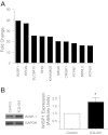
Neutrophil transmigration or β-catenin/p300 signaling induces Wnt-induced secreted protein (WISP1) expression in lung epithelial cells. A: neutrophils were induced to migrate across Calu-3 cells for 90 min followed by incubation in media for 4 h. Epithelial cell cDNA from 4 separate experiments was pooled and analyzed using a whole genome expression microarray (Agilent). The 10 most highly upregulated genes are shown. B: Calu-3 cells were treated with ICG-001 10 μM for 24 h and lysed. Cell lysates were analyzed by SDS-PAGE and immunoblotting for WISP1 and GAPDH. WISP1 protein levels were calculated relative to GAPDH levels by densitometry.
Fig. 6.
Fig. 6.
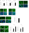
Neutrophil transmigration results in upregulation of WISP1 and cysteine-rich 61 (Cyr61) in lung epithelial cells. Neutrophils (polymorphonuclear leukocytes, PMN) were induced by a gradient of fMLP (1 μM) to migrate for 90 min across Calu-3 cells (A, C, and E) or primary human small airway epithelial cells (SAEC) (B and D), or Calu-3 cells (F) transfected with a nonsilencing shRNA or shRNA to β-catenin. E: ICG-001 (10 μM) was added to the monolayer after migration. A and B: real-time quantitative PCR analysis of WISP1 or Cyr61 expression was performed on epithelial cDNA isolated 2 h after the end of migration. CF: epithelial monolayers were fixed 28 h after migration and immunostained for WISP1 or Cyr61. E: fluorescence intensity was analyzed in the epithelial cells contiguous (C) to the denuded area and distant (D) from the sites of injury using ImageJ software. Arrows indicated enhanced epithelial WISP1 or Cyr61 expression at the sites of injury. *P ≤ 0.05, n ≥ 4. Error bars represent SE.
Fig. 7.
Fig. 7.
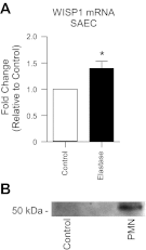
Neutrophil elastase triggers WISP1 upregulation and neutrophil transmigration results in cleavage of E-cadherin from the epithelial cell surface. A: primary human SAEC were treated with 0.1 U/ml purified elastase for 1 h and then incubated in media for 2 h. Epithelial cDNA was analyzed by qPCR for WISP1. B: neutrophils were induced to migrate across SAEC for 90 min. Epithelial cell supernatants were harvested from the apical surface, concentrated by ultracentrifugation through a 10-kDa MW filter, separated by SDS/PAGE, and immunoblotted with an antibody that recognizes the extracellular domain of E-cadherin (DECMA-1). *P ≤ 0.05, n ≥ 4.
Fig. 8.
Fig. 8.
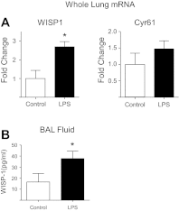
Neutrophilic inflammation is associated with upregulation of WISP1 in animal models. C57BL/6 mice were treated with LPS 20 μg in 50 μl saline or 50 μl of saline and euthanized at 4–5 days. A: lungs were snap frozen, and RNA was extracted, reverse transcribed to cDNA, and subjected to qPCR using primers for WISP1 and Cyr61. B: WISP1 ELISA was performed on BAL fluid. *P ≤ 0.05. Error bars represent SE. Difference in Cyr61 mRNA expression was not significant between control and LPS-treated lungs.
Fig. 9.
Fig. 9.
WISP1 and Cyr61 accelerate repair of the lung epithelium after inflammatory injury. Neutrophils were induced to migrate across monolayers of Calu-3-GFP cells. At 24 h after migration, epithelial monolayers were treated with rhWISP1 (A), rhCyr61 (B), or Cyr61-neutralizing antibody (C), as described in materials and methods. A: fluorescent images (×2.5) were acquired at the specified time points and the total cross-sectional area of epithelial defects in 5 HPF was measured. Area is normalized to control in each experiment. Arrowheads indicate epithelial wounds. B and C: TER was measured. In these experiments, the media were changed at 24 h after migration to remove secreted growth factors, which likely prevented the TER from returning to baseline in the control samples. *P < 0.05; n ≥ 4. Error bars represent SE.

Comment in

  • β-Catenin and CCNs in lung epithelial repair.
    Lawson WE, Blackwell TS. Lawson WE, et al. Am J Physiol Lung Cell Mol Physiol. 2013 May 1;304(9):L579-81. doi: 10.1152/ajplung.00073.2013. Epub 2013 Mar 22. Am J Physiol Lung Cell Mol Physiol. 2013. PMID: 23525784 Free PMC article. No abstract available.

Similar articles

Cited by

References

    1. Aarbiou J, Ertmann M, van Wetering S, van Noort P, Rook D, Rabe KF, Litvinov SV, van Krieken JH, de Boer WI, Hiemstra PS. Human neutrophil defensins induce lung epithelial cell proliferation in vitro. J Leukoc Biol 72: 167–174, 2002 - PubMed
    1. Adamson A, Perkins S, Brambilla E, Tripp S, Holden J, Travis W, Guinee D., Jr Proliferation, C-myc, and cyclin D1 expression in diffuse alveolar damage: potential roles in pathogenesis and implications for prognosis. Hum Pathol 30: 1050–1057, 1999 - PubMed
    1. Adamson IY, Bowden DH. The type 2 cell as progenitor of alveolar epithelial regeneration. A cytodynamic study in mice after exposure to oxygen. Lab Invest 30: 35–42, 1974 - PubMed
    1. Ashino Y, Ying X, Dobbs LG, Bhattacharya J. [Ca(2+)](i) oscillations regulate type II cell exocytosis in the pulmonary alveolus. Am J Physiol Lung Cell Mol Physiol 279: L5–L13, 2000 - PubMed
    1. Atabai K, Ishigaki M, Geiser T, Ueki I, Matthay MA, Ware LB. Keratinocyte growth factor can enhance alveolar epithelial repair by nonmitogenic mechanisms. Am J Physiol Lung Cell Mol Physiol 283: L163–L169, 2002 - PubMed

Publication types

MeSH terms

LinkOut - more resources