. 2013 Jan 3:6:114.
doi: 10.3389/fncir.2012.00114. eCollection 2012.

Cortical modulation of auditory processing in the midbrain

Affiliations

Cortical modulation of auditory processing in the midbrain

Victoria M Bajo et al. Front Neural Circuits. .

Abstract

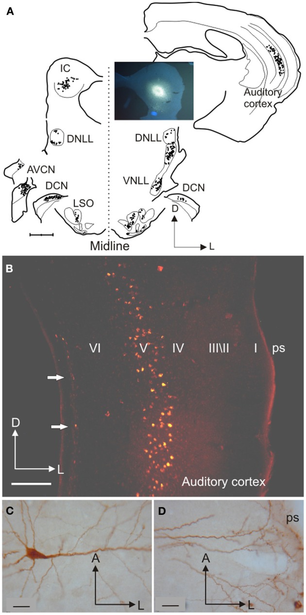
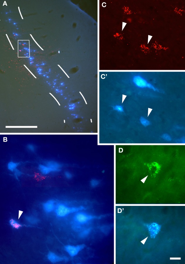
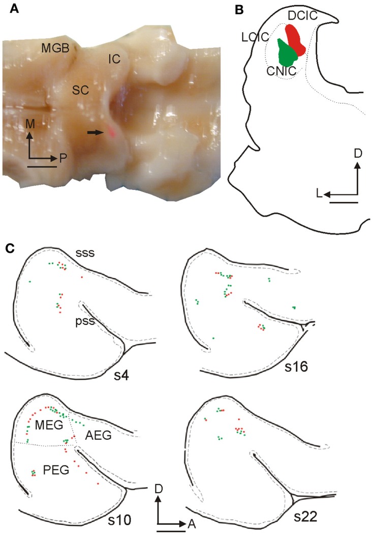
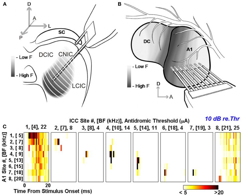
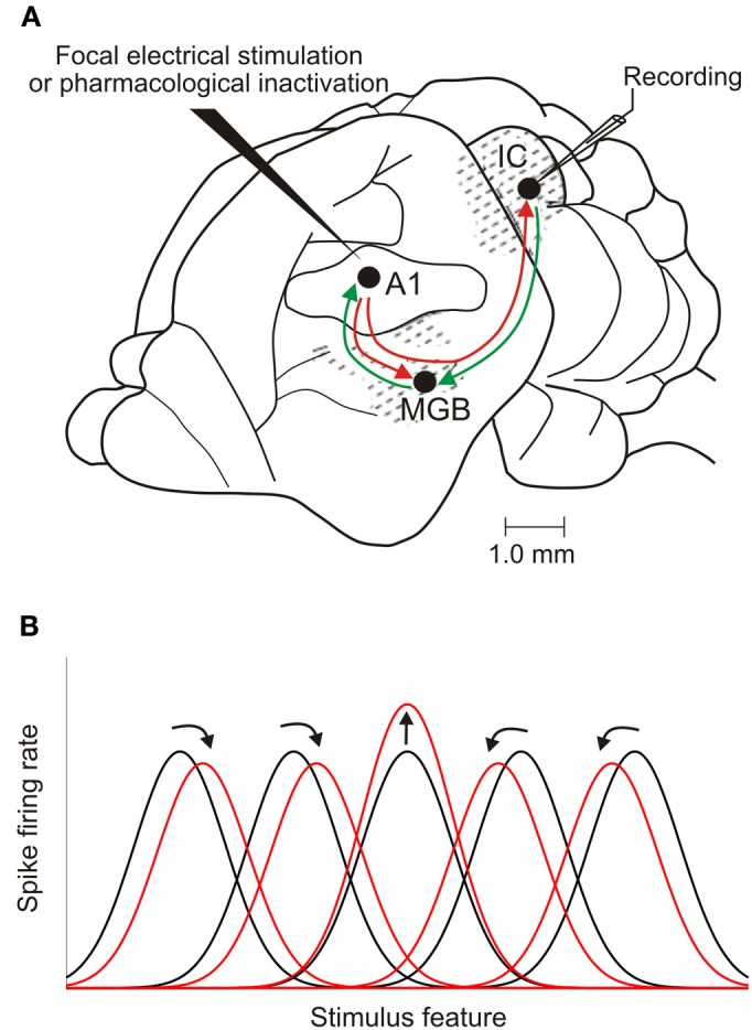
In addition to their ascending pathways that originate at the receptor cells, all sensory systems are characterized by extensive descending projections. Although the size of these connections often outweighs those that carry information in the ascending auditory pathway, we still have a relatively poor understanding of the role they play in sensory processing. In the auditory system one of the main corticofugal projections links layer V pyramidal neurons with the inferior colliculus (IC) in the midbrain. All auditory cortical fields contribute to this projection, with the primary areas providing the largest outputs to the IC. In addition to medium and large pyramidal cells in layer V, a variety of cell types in layer VI make a small contribution to the ipsilateral corticocollicular projection. Cortical neurons innervate the three IC subdivisions bilaterally, although the contralateral projection is relatively small. The dorsal and lateral cortices of the IC are the principal targets of corticocollicular axons, but input to the central nucleus has also been described in some studies and is distinctive in its laminar topographic organization. Focal electrical stimulation and inactivation studies have shown that the auditory cortex can modify almost every aspect of the response properties of IC neurons, including their sensitivity to sound frequency, intensity, and location. Along with other descending pathways in the auditory system, the corticocollicular projection appears to continually modulate the processing of acoustical signals at subcortical levels. In particular, there is growing evidence that these circuits play a critical role in the plasticity of neural processing that underlies the effects of learning and experience on auditory perception by enabling changes in cortical response properties to spread to subcortical nuclei.

Keywords: auditory cortex; corticofugal; descending projection; inferior colliculus; plasticity; sound localization.


Figures

Figure 1
Figure 1
Inputs from different auditory centers converge in the IC. (A) A small fluorogold tracer injection in the ventromedial part of the IC central nucleus of the gerbil produces retrograde labeling of neurons in the MSO, periolivary nuclei, VNLL, and A1 on the same side, in the cochlear nuclei and IC on the opposite side, and in the LSO, DCN, and DNLL on both sides. (B) Retrogradely labeled cells in the cortex are found mainly in layer V after a rhodamine tracer injection in the IC. (C) Large labeled pyramidal cell with the soma located in cortical layer V and (D) a tufted dendritic tree ending in layer I. Calibration bars: 1 mm (A), 0.2 mm (B), and 25 μm (C,D). Modified with permission from Bajo and Moore (2005).
Figure 2
Figure 2
Anatomical tracing experiments in guinea pigs show that cortical cells project to the IC bilaterally. (A) Coronal section at the level of the right auditory cortex showing the overlay of cells labeled retrogradely by injections of Fast Blue in the right IC and rhodamine microbeads in the left IC, with a higher magnification of the area enclosed by the white box in (B). (C,C') Double-labeled cortical cells (arrowheads) using the same tracer combination. (D,D') A double-labeled cortical cell following an injection of Fast Blue in the ipsilateral IC and fluorescein microbeads in the contralateral IC. Arrowheads indicate double labeled cells. Calibration bars: 0.5 mm (A) and 20 μm (B–D). Modified with permission from Coomes et al. (2005).
Figure 3
Figure 3
Retrogradely labeled cells in ferret auditory cortex after fluorescent microbead injections in the IC. (A) Dorsal view of the ferret brain where both the cerebral cortices and the cerebellum were removed to visualize the thalamus and midbrain. A rhodamine microbead injection site can be seen in the left IC (arrow). (B) Coronal section at the level of the IC from this animal illustrating rhodamine and fluorescein microbead injection sites. (C) Drawings of tangential 50 μm sections spaced 300 μm apart from lateral to medial (s22 is the most medial) at the level of the left ectosylvian gyrus where the auditory cortex is located, showing green and red retrogradely labeled cells. Calibration bars: 2 mm (A,C) and 1 mm (B). Based on Bajo et al. (2007).
Figure 4
Figure 4
Terminal fields in the IC after a tracer injection in the ferret auditory cortex. (A) Coronal section at the level of the left auditory cortex showing the location of a rhodamine injection site in the center of the MEG where A1 is located. The halo of the injection site is shown in gray while the center is in black. (B–D) Examples of anterograde terminal fields in the IC at the locations indicated by the boxes in (E). Calibration bars: 1 mm (A), 100 μm (B–D), 0.5 mm (E). Modified with permission from Bajo et al. (2007).
Figure 5
Figure 5
Electron micrographs of labeled endings in the three main subdivisions of the IC after a large injection of biotinylated dextran amine was made in the ipsilateral primary auditory cortex in the rat. Labeled terminals in DCIC (A), CNIC (B), and LCIC (C) contain round vesicles and make asymmetric synaptic contacts (arrows). Unlabeled terminals with pleomorphic vesicles are also observed (stars). Inset in panel (A) shows the distribution of corticocollicular terminal fields and the areas (the box in each IC subdivision) that were used for electron microscopy. Calibration bar: 0.4 μm. Modified with permission from Saldaña et al. (1996).
Figure 6
Figure 6
Antidromic activation reveals tonotopically organized projections from A1 to the CNIC in guinea pig. Multi-site probes were positioned along the tonotopic axis of the guinea pig CNIC (A) and A1 (B). (C) Post-stimulus time histograms (PSTHs) for eight A1 locations with different best frequencies (BF) from low (location 1) to high (location 8). These responses were evoked by antidromic stimulation equivalent to 10 dB above threshold at eight frequency-matched locations in the IC. Color scale corresponds to the total number of spikes across 40 trials where any values ≤5 and ≥20 were set to white and black, respectively. Modified with permission from Lim and Anderson (2007).
Figure 7
Figure 7
Corticofugal modulation of IC response properties. (A) Lateral view of the brain of a mustached bat, one of the species used most in cortical stimulation and inactivation experiments. The auditory cortex has reciprocal ascending and descending connections with the medial geniculate body (MGB) in the thalamus. It also sends a descending projection to the IC, which, in turn, projects to the MGB. (B) Focal electrical stimulation in the cortex results in facilitation of the responses of IC neurons that have tuning properties matched to those of cortical neurons at the site of the stimulating electrode. The tuning of unmatched IC neurons may shift toward that of the stimulated cortical neurons (as illustrated here), resulting in an expanded representation of the stimulus feature. Shifts in tuning away from that of the stimulated cortical neurons have also been described, compressing the midbrain representation. Adapted with permission from Suga (2012).
Figure 8
Figure 8
Inactivation of the auditory cortex alters sensitivity to interaural level differences (ILD) in the inferior colliculus. (A) Schematic of the experimental setup, showing a cooling probe (blue) above the auditory cortex and a recording electrode in the ipsilateral IC. (B) Examples of rate-ILD functions obtained before, during and after cortical inactivation. Adapted with permission from Nakamoto et al. (2008).
Figure 9
Figure 9
The projection from the auditory cortex to the inferior colliculus is essential for training-induced plasticity of spatial hearing in adult ferrets. (A) Lateral view of the ferret midbrain showing the location and number of injections of fluorescent microspheres conjugated with chlorine e6 in the left IC. (B) Schematic showing the selective ablation of retrogradely labeled layer V corticocollicular neurons by illumination of the ipsilateral auditory cortex with near-infrared light. (C) Sound localization accuracy (averaged across 12 speaker locations in the horizontal plane) before the right ear was plugged (Preplug), on each of the 10 days over which an ear plug was worn and following its removal (Post-plug). Data from control animals are shown in black and from the ferrets with corticocollicular lesions in red. The symbols represent different animals and the lines show the mean scores. (D) Staining with the SMI32 antibody, a marker of layer III and layer V pyramidal cortical neurons, was sparser in the left (lesioned) primary auditory cortex, resulting in a less distinct bilaminar appearance (top) than on the right side (bottom). Calibration bars: 2 mm in (A) and 0.1 mm in (D). Modified with permission from Bajo et al. (2010a).

Similar articles

Cited by

References

    1. Aitkin L. M., Phillips S. C. (1984). Is the inferior colliculus an obligatory relay in the cat auditory system? Neurosci. Lett. 44, 259–264 - PubMed
    1. Andersen R. A., Snyder R. L., Merzenich M. M. (1980). The topographic organization of corticocollicular projections from physiologically identified loci in the AI, AII, and anterior auditory cortical fields of the cat. J. Comp. Neurol. 191, 479–494 10.1002/cne.901910310 - DOI - PubMed
    1. Bajo V. M., Moore D. R. (2005). Descending projections from the auditory cortex to the inferior colliculus in the gerbil, Meriones unguiculatus. J. Comp. Neurol. 486, 101–116 10.1002/cne.20542 - DOI - PubMed
    1. Bajo V. M., Nodal F. R., Bizley J. K., Moore D. R., King A. J. (2007). The ferret auditory cortex: descending projections to the inferior colliculus. Cereb. Cortex 17, 475–491 10.1093/cercor/bhj164 - DOI - PMC - PubMed
    1. Bajo V. M., Nodal F. R., Moore D. R., King A. J. (2010a). The descending corticocollicular pathway mediates learning-induced auditory plasticity. Nat. Neurosci. 13, 253–260 10.1038/nn.2466 - DOI - PMC - PubMed

LinkOut - more resources