

The Transcriptional Response of Neurotrophins and Their Tyrosine Kinase Receptors in Lumbar Sensorimotor Circuits to Spinal Cord Contusion is Affected by Injury Severity and Survival Time
- PMID: 23316162
- PMCID: PMC3540763
- DOI: 10.3389/fphys.2012.00478
The Transcriptional Response of Neurotrophins and Their Tyrosine Kinase Receptors in Lumbar Sensorimotor Circuits to Spinal Cord Contusion is Affected by Injury Severity and Survival Time
Abstract
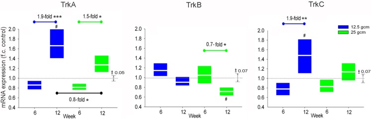
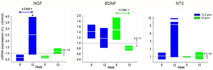
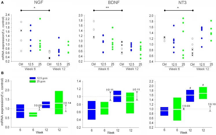
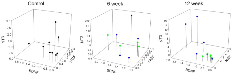
Traumatic spinal cord injury (SCI) results in changes to the anatomical, neurochemical, and physiological properties of cells in the central and peripheral nervous system. Neurotrophins, acting by binding to their cognate Trk receptors on target cell membranes, contribute to modulation of anatomical, neurochemical, and physiological properties of neurons in sensorimotor circuits in both the intact and injured spinal cord. Neurotrophin signaling is associated with many post-SCI changes including maladaptive plasticity leading to pain and autonomic dysreflexia, but also therapeutic approaches such as training-induced locomotor improvement. Here we characterize expression of mRNA for neurotrophins and Trk receptors in lumbar dorsal root ganglia (DRG) and spinal cord after two different severities of mid-thoracic injury and at 6 and 12 weeks post-SCI. There was complex regulation that differed with tissue, injury severity, and survival time, including reversals of regulation between 6 and 12 weeks, and the data suggest that natural regulation of neurotrophins in the spinal cord may continue for months after birth. Our assessments determined that a coordination of gene expression emerged at the 12-week post-SCI time point and bioinformatic analyses address possible mechanisms. These data can inform studies meant to determine the role of the neurotrophin signaling system in post-SCI function and plasticity, and studies using this signaling system as a therapeutic approach.
Keywords: contusions; genetic regulation; injury mechanisms; neurotrophin receptors; neurotrophins; sensory neurons; spinal cord injury; transcription.
Figures


Similar articles
-
Comparisons of motor and sensory abnormalities after lumbar and thoracic contusion spinal cord injury in male rats.Neurosci Lett. 2019 Aug 24;708:134358. doi: 10.1016/j.neulet.2019.134358. Epub 2019 Jun 30. Neurosci Lett. 2019. PMID: 31269465
-
miR-155 Deletion in Mice Overcomes Neuron-Intrinsic and Neuron-Extrinsic Barriers to Spinal Cord Repair.J Neurosci. 2016 Aug 10;36(32):8516-32. doi: 10.1523/JNEUROSCI.0735-16.2016. J Neurosci. 2016. PMID: 27511021 Free PMC article.
-
Upregulation of calcium channel alpha-2-delta-1 subunit in dorsal horn contributes to spinal cord injury-induced tactile allodynia.Spine J. 2018 Jun;18(6):1062-1069. doi: 10.1016/j.spinee.2018.01.010. Epub 2018 Jan 31. Spine J. 2018. PMID: 29355786
-
Role of neurotrophins in spinal plasticity and locomotion.Curr Pharm Des. 2013;19(24):4509-16. doi: 10.2174/13816128113199990378. Curr Pharm Des. 2013. PMID: 23360280 Review.
-
Transplants and neurotrophic factors increase regeneration and recovery of function after spinal cord injury.Prog Brain Res. 2002;137:257-73. doi: 10.1016/s0079-6123(02)37020-1. Prog Brain Res. 2002. PMID: 12440372 Review.
Cited by
-
Analysis of the Expression of Neurotrophins and Their Receptors in Adult Zebrafish Kidney.Vet Sci. 2022 Jun 15;9(6):296. doi: 10.3390/vetsci9060296. Vet Sci. 2022. PMID: 35737348 Free PMC article.
-
TrkB Agonist (7,8-DHF)-Induced Responses in Dorsal Root Ganglia Neurons Are Decreased after Spinal Cord Injury: Implication for Peripheral Pain Mechanisms.eNeuro. 2025 Jan 3;12(1):ENEURO.0219-24.2024. doi: 10.1523/ENEURO.0219-24.2024. Print 2025 Jan. eNeuro. 2025. PMID: 39753357 Free PMC article.
-
Drug delivery, cell-based therapies, and tissue engineering approaches for spinal cord injury.J Control Release. 2015 Dec 10;219:141-154. doi: 10.1016/j.jconrel.2015.08.060. Epub 2015 Sep 4. J Control Release. 2015. PMID: 26343846 Free PMC article. Review.
-
Challenges and opportunities of sensory plasticity after SCI.Front Physiol. 2013 Aug 27;4:231. doi: 10.3389/fphys.2013.00231. eCollection 2013. Front Physiol. 2013. PMID: 23986722 Free PMC article. No abstract available.
-
Novel multi-system functional gains via task specific training in spinal cord injured male rats.J Neurotrauma. 2014 May 1;31(9):819-33. doi: 10.1089/neu.2013.3082. Epub 2014 Mar 25. J Neurotrauma. 2014. PMID: 24294909 Free PMC article.
References
-
- Allen Institute for Brain Science (2009). Allen Spinal Cord Atlas [Internet]. Seattle, WA: Available at: http://mousespinal.brain-map.org
-
- Arvanian B. W., VI, Anderson A., Horner P. J., Federoff H. J., Mendell L. M. (2006a). Combined delivery of neurotrophin-3 and NMDA receptors 2D subunit strengthens synaptic transmission in contused and staggered double hemisected spinal cord of neonatal rat. Exp. Neurol. 197, 347–35210.1016/j.expneurol.2005.10.008 - DOI - PubMed
Grants and funding
LinkOut - more resources
Full Text Sources
Other Literature Sources