

The "sweet" side of a long pentraxin: how glycosylation affects PTX3 functions in innate immunity and inflammation
- PMID: 23316195
- PMCID: PMC3539679
- DOI: 10.3389/fimmu.2012.00407
The "sweet" side of a long pentraxin: how glycosylation affects PTX3 functions in innate immunity and inflammation
Abstract
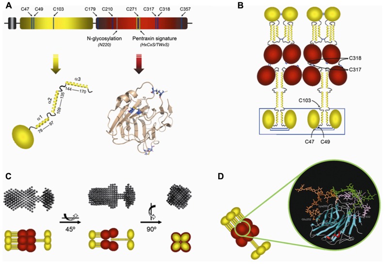
Innate immunity represents the first line of defense against pathogens and plays key roles in activation and orientation of the adaptive immune response. The innate immune system comprises both a cellular and a humoral arm. Components of the humoral arm include soluble pattern recognition molecules (PRMs) that recognize pathogen-associated molecular patterns and initiate the immune response in coordination with the cellular arm, therefore acting as functional ancestors of antibodies. The long pentraxin PTX3 is a prototypic soluble PRM that is produced at sites of infection and inflammation by both somatic and immune cells. Gene targeting of this evolutionarily conserved protein has revealed a non-redundant role in resistance to selected pathogens. Moreover, PTX3 exerts important functions at the crossroad between innate immunity, inflammation, and female fertility. The human PTX3 protein contains a single N-glycosylation site that is fully occupied by complex type oligosaccharides, mainly fucosylated and sialylated biantennary glycans. Glycosylation has been implicated in a number of PTX3 activities, including neutralization of influenza viruses, modulation of the complement system, and attenuation of leukocyte recruitment. Therefore, this post translational modification might act as a fine tuner of PTX3 functions in native immunity and inflammation. Here we review the studies on PTX3, with emphasis on the glycan-dependent mechanisms underlying pathogen recognition and crosstalk with other components of the innate immune system.
Keywords: PTX3; glycosylation; inflammation; pathogen recognition; pentraxins.
Figures

Similar articles
-
Pentraxins in humoral innate immunity.Adv Exp Med Biol. 2012;946:1-20. doi: 10.1007/978-1-4614-0106-3_1. Adv Exp Med Biol. 2012. PMID: 21948359 Review.
-
Pathogen recognition by the long pentraxin PTX3.J Biomed Biotechnol. 2011;2011:830421. doi: 10.1155/2011/830421. Epub 2011 Jun 2. J Biomed Biotechnol. 2011. PMID: 21716666 Free PMC article. Review.
-
The long pentraxin PTX3 at the crossroads between innate immunity and tissue remodelling.Tissue Antigens. 2011 Apr;77(4):271-82. doi: 10.1111/j.1399-0039.2011.01645.x. Tissue Antigens. 2011. PMID: 21388349
-
The long pentraxin PTX3 as a key component of humoral innate immunity and a candidate diagnostic for inflammatory diseases.Int Arch Allergy Immunol. 2014;165(3):165-78. doi: 10.1159/000368778. Epub 2014 Dec 20. Int Arch Allergy Immunol. 2014. PMID: 25531094 Review.
-
The long pentraxin PTX3: a paradigm for humoral pattern recognition molecules.Ann N Y Acad Sci. 2013 May;1285:1-14. doi: 10.1111/nyas.12043. Epub 2013 Mar 25. Ann N Y Acad Sci. 2013. PMID: 23527487 Review.
Cited by
-
Targeting chemotherapy-induced PTX3 in tumor stroma to prevent the progression of drug-resistant cancers.Oncotarget. 2015 Sep 15;6(27):23987-4001. doi: 10.18632/oncotarget.4364. Oncotarget. 2015. PMID: 26124179 Free PMC article.
-
Pentraxin-3-mediated complement activation in a swine model of renal ischemia/reperfusion injury.Aging (Albany NY). 2021 Apr 20;13(8):10920-10933. doi: 10.18632/aging.202992. Epub 2021 Apr 20. Aging (Albany NY). 2021. PMID: 33875620 Free PMC article.
-
Maternal serum pentraxin 3 level in early pregnancy for prediction of gestational diabetes mellitus.Ann Transl Med. 2019 Dec;7(23):722. doi: 10.21037/atm.2019.12.25. Ann Transl Med. 2019. PMID: 32042738 Free PMC article.
-
Control of Complement Activation by the Long Pentraxin PTX3: Implications in Age-Related Macular Degeneration.Front Pharmacol. 2020 Nov 26;11:591908. doi: 10.3389/fphar.2020.591908. eCollection 2020. Front Pharmacol. 2020. PMID: 33324220 Free PMC article.
-
C1QL3 promotes cell-cell adhesion by mediating complex formation between ADGRB3/BAI3 and neuronal pentraxins.FASEB J. 2021 Jan;35(1):e21194. doi: 10.1096/fj.202000351RR. FASEB J. 2021. PMID: 33337553 Free PMC article.
References
-
- Abernethy T. J., Avery O. T. (1941). The occurrence during acute infections of a protein non normally present in the blood. I. Distribution of the reactive protein in patients’ sera and the effect of calcium on the flocculation reaction with C. Polysaccharide of pneumococcus. J. Exp. Med. 73 173–182 - PMC - PubMed
-
- Akira S., Uematsu S., Takeuchi O. (2006). Pathogen recognition and innate immunity. Cell 124 783–801 - PubMed
-
- Altmeyer A., Klampfer L., Goodman A. R., Vilcek J. (1995). Promoter structure and transcriptional activation of the murine TSG-14 gene encoding a tumor necrosis factor/interleukin-1-inducible pentraxin protein. J. Biol. Chem. 270 25584–25590 - PubMed
-
- Anders E. M., Hartley C. A., Reading P. C., Ezekowitz R. A. (1994). Complement-dependent neutralization of influenza virus by a serum mannose-binding lectin. J. Gen. Virol. 75(Pt 3) 615–622 - PubMed
-
- Andersen O., Vilsgaard Ravn K., Juul Sorensen I., Jonson G., Holm Nielsen E., Svehag S. E. (1997). Serum amyloid P component binds to influenza A virus haemagglutinin and inhibits the virus infection in vitro. Scand. J. Immunol. 46 331–337 - PubMed
LinkOut - more resources
Full Text Sources
Other Literature Sources
Miscellaneous