

Fundus autofluorescence imaging in an ocular screening program
- PMID: 23316224
- PMCID: PMC3536047
- DOI: 10.1155/2012/806464
Fundus autofluorescence imaging in an ocular screening program
Abstract
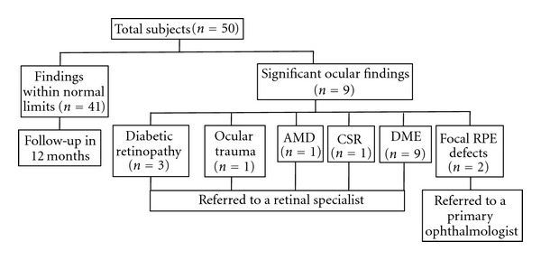
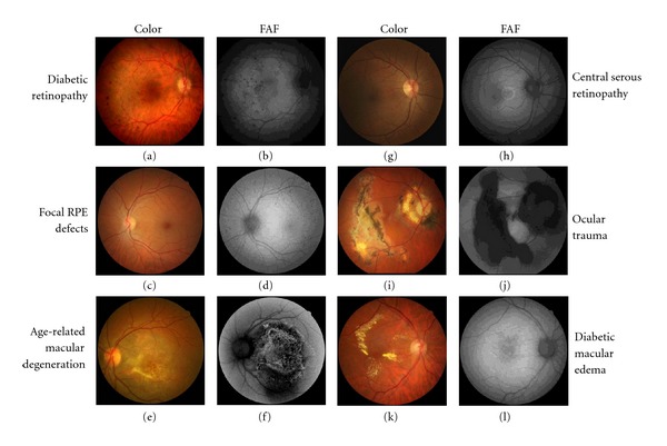
Purpose. To describe integration of fundus autofluorescence (FAF) imaging into an ocular screening program. Methods. Fifty consecutive screening participants were included in this prospective pilot imaging study. Color and FAF (530/640 nm exciter/barrier filters) images were obtained with a 15.1MP Canon nonmydriatic hybrid camera. A clinician evaluated the images on site to determine need for referral. Visual acuity (VA), intraocular pressure (IOP), and ocular pathology detected by color fundus and FAF imaging modalities were recorded. Results. Mean ± SD age was 47.4 ± 17.3 years. Fifty-two percent were female and 58% African American. Twenty-seven percent had a comprehensive ocular examination within the past year. Mean VA was 20/39 in the right eye and 20/40 in the left eye. Mean IOP was 15 mmHg bilaterally. Positive color and/or FAF findings were identified in nine (18%) individuals with diabetic retinopathy or macular edema (n = 4), focal RPE defects (n = 2), age-related macular degeneration (n = 1), central serous retinopathy (n = 1), and ocular trauma (n = 1). Conclusions. FAF was successfully integrated in our ocular screening program and aided in the identification of ocular pathology. Larger studies examining the utility of this technology in screening programs may be warranted.
Figures
Similar articles
-
Fundus autofluorescence and colour fundus imaging compared during telemedicine screening in patients with diabetes.J Telemed Telecare. 2013 Jun;19(4):209-12. doi: 10.1177/1357633x13492292. Epub 2013 Jun 14. J Telemed Telecare. 2013. PMID: 24163061
-
Correlation of fundus autofluorescence with spectral-domain optical coherence tomography and vision in diabetic macular edema.Ophthalmology. 2012 May;119(5):1056-65. doi: 10.1016/j.ophtha.2011.11.018. Epub 2012 Feb 18. Ophthalmology. 2012. PMID: 22342014
-
Two-wavelength fundus autofluorescence and macular pigment optical density imaging in diabetic macular oedema.Eye (Lond). 2012 Aug;26(8):1078-85. doi: 10.1038/eye.2012.100. Epub 2012 Jun 15. Eye (Lond). 2012. PMID: 22699976 Free PMC article.
-
Spotlight on fundus autofluorescence.Clin Optom (Auckl). 2018 Mar 27;10:25-32. doi: 10.2147/OPTO.S134637. eCollection 2018. Clin Optom (Auckl). 2018. PMID: 30214339 Free PMC article. Review.
-
Novel imaging biomarkers in diabetic retinopathy and diabetic macular edema.Ther Adv Ophthalmol. 2020 Sep 4;12:2515841420950513. doi: 10.1177/2515841420950513. eCollection 2020 Jan-Dec. Ther Adv Ophthalmol. 2020. PMID: 32954207 Free PMC article. Review.
Cited by
-
Fundus Autofluorescence Captured With a Nonmydriatic Retinal Camera in Vegetarians Versus Nonvegetarians.J Diabetes Sci Technol. 2015 Sep 9;10(1):151-6. doi: 10.1177/1932296815599003. J Diabetes Sci Technol. 2015. PMID: 26353779 Free PMC article.
-
Pilot Study on Visual Function and Fundus Autofluorescence Assessment in Diabetic Patients.J Ophthalmol. 2016;2016:1287847. doi: 10.1155/2016/1287847. Epub 2016 Feb 10. J Ophthalmol. 2016. PMID: 26977312 Free PMC article.
-
Long-term development of lens fluorescence in a twin cohort: Heritability and effects of age and lifestyle.PLoS One. 2022 May 26;17(5):e0268458. doi: 10.1371/journal.pone.0268458. eCollection 2022. PLoS One. 2022. PMID: 35617652 Free PMC article.
References
-
- Yogesan K, Constable IJ, Eikelboom RH, van Saarloos PP. Tele-ophthalmic screening using digital imaging devices. Australian and New Zealand Journal of Ophthalmology. 1998;26(supplement 1):S9–S11. - PubMed
-
- Khouri AS, Szirth BC, Salti HI, Fechtner RD. DICOM transmission of simultaneous stereoscopic images of the optic nerve in patients with glaucoma. Journal of Telemedicine and Telecare. 2007;13(7):337–340. - PubMed
-
- Pirbhai A, Sheidow T, Hooper P. Prospective evaluation of digital non-stereo color fundus photography as a screening tool in age-related macular degeneration. American Journal of Ophthalmology. 2005;139(3):455–461. - PubMed
-
- Khouri AS, Szirth BC, Shahid KS, Fechtner RD. Software-assisted optic nerve assessment for glaucoma tele-screening. Telemedicine and e-Health. 2008;14(3):261–265. - PubMed
-
- Tanabe N, Go K, Sakurada Y, et al. A remote operating slit lamp microscope system. Development and its utility in ophthalmologic examinations. Methods of Information in Medicine. 2011;50:427–434. - PubMed
LinkOut - more resources
Full Text Sources
Miscellaneous
