Comparative Study
. 2012 Dec;1(6):e005652.
doi: 10.1161/JAHA.112.005652. Epub 2012 Dec 19.

In vivo cardiac cellular reprogramming efficacy is enhanced by angiogenic preconditioning of the infarcted myocardium with vascular endothelial growth factor

Affiliations
Comparative Study

In vivo cardiac cellular reprogramming efficacy is enhanced by angiogenic preconditioning of the infarcted myocardium with vascular endothelial growth factor

Megumi Mathison et al. J Am Heart Assoc. 2012 Dec.

Abstract

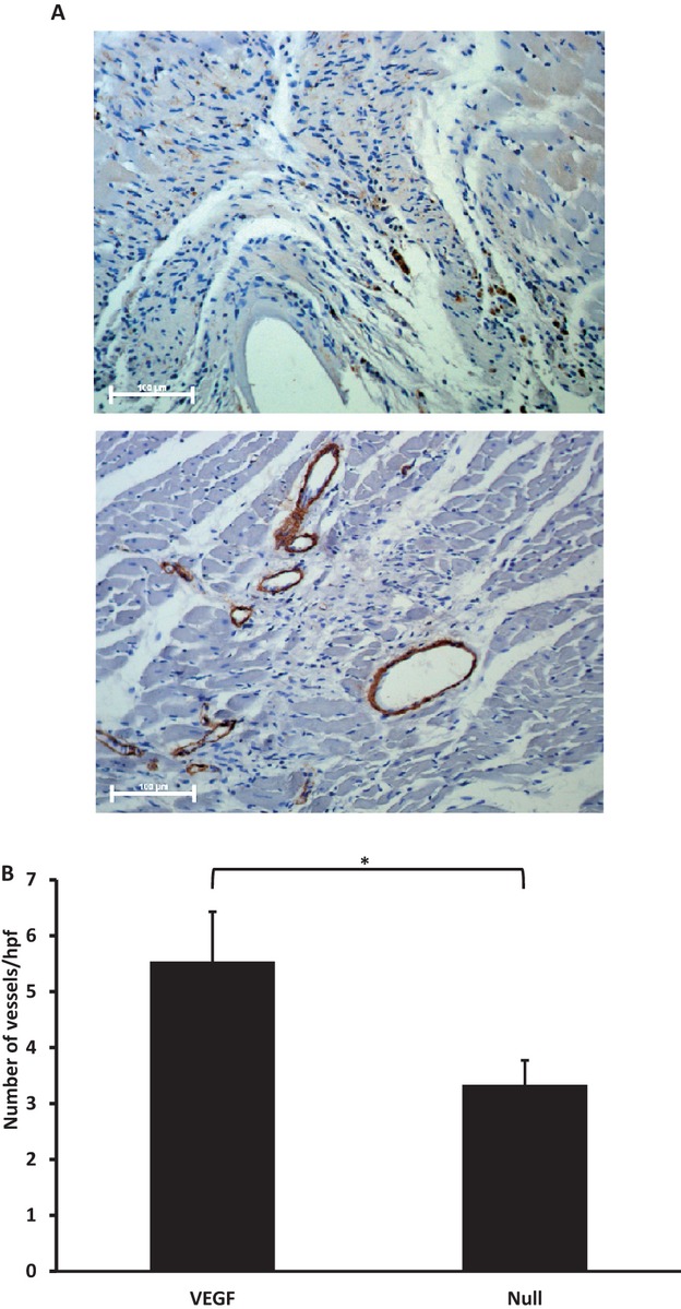
Background: In situ cellular reprogramming offers the possibility of regenerating functional cardiomyocytes directly from scar fibroblasts, obviating the challenges of cell implantation. We hypothesized that pretreating scar with gene transfer of the angiogenic vascular endothelial growth factor (VEGF) would enhance the efficacy of this strategy.

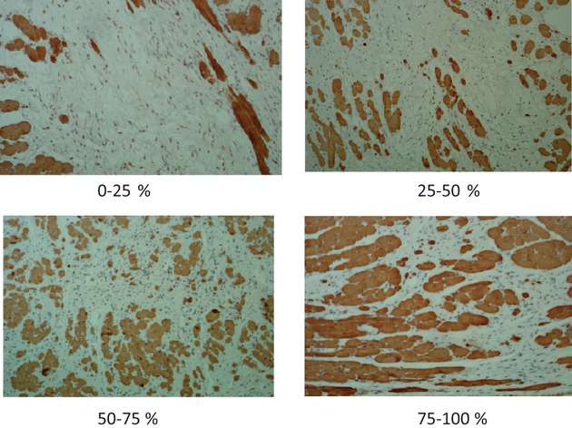
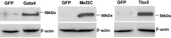
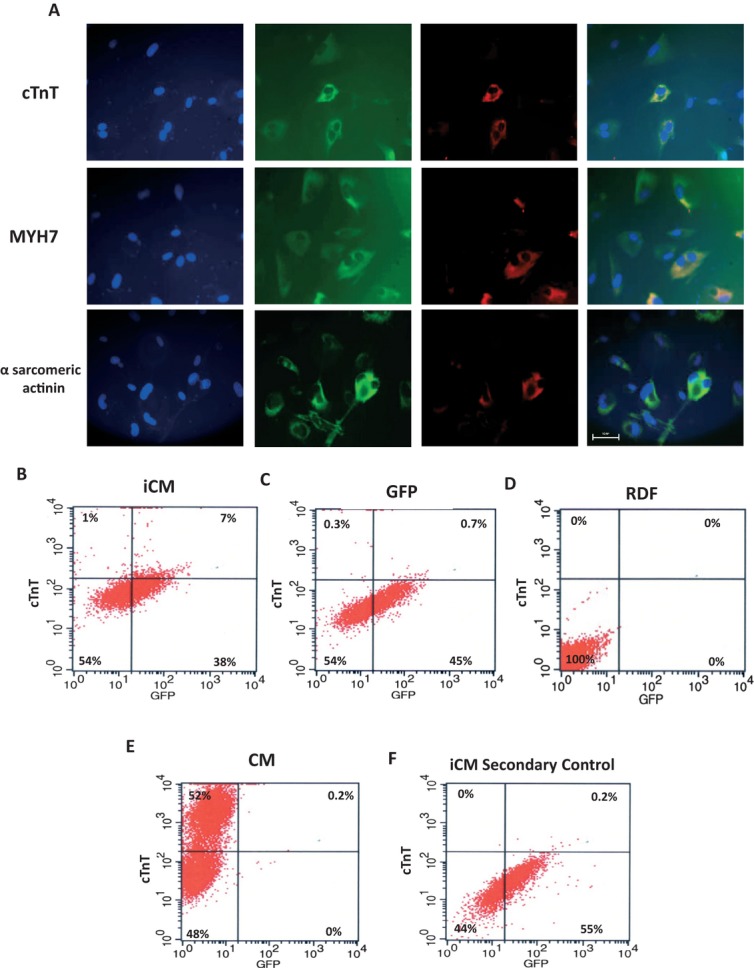
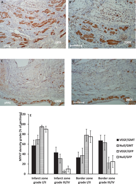
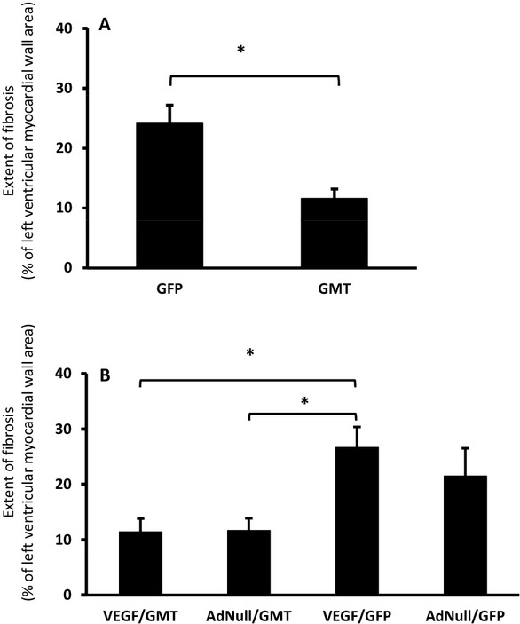
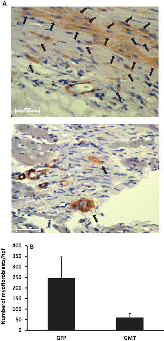
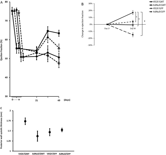
Methods and results: Gata4, Mef2c, and Tbx5 (GMT) administration via lentiviral transduction was demonstrated to transdifferentiate rat fibroblasts into (induced) cardiomyocytes in vitro by cardiomyocyte marker studies. Fisher 344 rats underwent coronary ligation and intramyocardial administration of an adenovirus encoding all 3 major isoforms of VEGF (AdVEGF-All6A(+)) or an AdNull control vector (n=12/group). Lentivirus encoding GMT or a GFP control was administered to each animal 3 weeks later, followed by histologic and echocardiographic analyses. GMT administration reduced the extent of fibrosis by half compared with GFP controls (12 ± 2% vs 24 ± 3%, P<0.01) and reduced the number of myofibroblasts detected in the infarct zone by 4-fold. GMT-treated animals also demonstrated greater density of cardiomyocyte-specific marker beta myosin heavy chain 7(+) cells compared with animals receiving GFP with or without VEGF (P<0.01). Ejection fraction was significantly improved after GMT vs GFP administration (12 ± 3% vs -7 ± 3%, P<0.01). Eight (73%) GFP animals but no GMT animals demonstrated decreased ejection fraction during this interval (P<0.01). Also, improvement in ejection fraction was 4-fold greater in GMT/VEGF vs GMT/null animals (17 ± 2% vs 4 ± 1%, P<0.05).

Conclusions: VEGF administration to infarcted myocardium enhances the efficacy of GMT-mediated cellular reprogramming in improving myocardial function and reducing the extent of myocardial fibrosis compared with the use of GMT or VEGF alone.

Keywords: angiogenesis; gene therapy; myocardial infarction; stem cells.


Figures

Figure 1.
Figure 1.
MHY7 cell density classification. MYH7+ cell number was analyzed in a semiquantitative manner as the MYH7+ cell density in 5 areas per slide viewed at ×200 magnification (at the center of the infarction zone, in the mid areas between the center of infarction and the border zone, and in each border zone adjacent to the infarct). These fields were graded by an investigator blinded to treatment group as follows: (top left) grade I: <25% of selected microscopic field demonstrating MYH7+ cells; (top right) grade II: 25% to 50% of selected microscopic field demonstrating MYH7+ cells; (bottom left) grade III: 50% to 75% of selected microscopic field demonstrating MYH7+ cells; and (bottom right) grade IV: >75% of selected microscopic field demonstrating MYH7+ cells. MYH7 indicates myosin heavy chain 7.
Figure 2.
Figure 2.
Generation of lentivirus encoding individual cardiac transcription factors. Lentivirus encoding the transcription factors Gata4, Mef2c, and Tbx5 were prepared in 293T package cells as described in Methods and Results and protein expression was detected by Western blot analysis using antibodies specific to each transgene. A lentivirus expressing a GFP marker gene was used as a control for these expression vectors and β‐actin was used as a loading control. GFP indicates green fluorescent protein.
Figure 3.
Figure 3.
iCM generation in vitro. Primary rat dermal fibroblast (RDF) cells were cultured and infected with GMT or a GFP control lentivirus as described in Methods and Results. Fourteen days after infection, cells were fixed and stained for specified cardiomyocyte markers. A, Immunofluorescence studies. First column represents 4',6‐diamidino‐2‐phenylindole (DAPI) staining to identify cell nuclei. Second column represents GFP fluorescence to identify cells infected by at least one of the lentivirus vectors. Third column represents red staining of relevant cardiomyocyte markers (first row: cardiac troponin T [cTnT]; second row: myosin heavy chain 7 [MHY7]; third row: α‐sarcomeric actinin). Fourth column depicts merge of previous 3 images. Note coincidence of these respective markers and binucleated cells typical of cardiomyocyte, and that GFP (−) cells also fail to express markers. Uninfected RDFs did not express either marker (not shown). All photomicrographs were taken at ×400 magnification (bar=50 μm). B through F, FACS analysis. Depicted are FACS plots for cTnT staining after: (B) RDFs infected with GMT, demonstrating 7% expression of cTnT in GFP+ cells, (C) RDFs infected with GFP control lentivirus, (D) uninfected RDFs, (E) primary cardiomyocyte control, and (F) RDFs infected with GMT, with use of secondary antibody only. Graphs show a minimum of 5000 events. iCM indicates induced cardiomyocytes; GMT, Gata4, Mef 2c, and Tbx5; FACS, fluorescence activated cell sorting.
Figure 4.
Figure 4.
Myocardial vascularization studies. Vascularization of infarct regions assessed by staining for α‐smooth muscle actin in sections obtained as described in Methods and Results 7 weeks after coronary ligation and administration of AdVEGF‐All6A+ or the control vector AdNull. A, Photomicrographs of representative sections of infarct zones viewed at ×200 after administration of AdNull/GMT (top) or AdVEGF‐All6A+/GMT (bottom). Bars represent 100 μm. B, Quantification of vascularization of infarct regions as assessed by the number of vessel per high power (×200) microscopic field (n=12/group). *P<0.05. VEGF indicates vascular endothelial growth factor; GMT, Gata4, Mef2c, and Tbx5.
Figure 5.
Figure 5.
Cardiomyocyte density in infarct zones. Cardiomyocyte‐specific marker MYH7 staining of the infarct and border zones of sections of myocardium harvested 7 weeks after coronary ligation and administration of AdVEGF‐All6A+ or the control vector AdNull (4 weeks after the administration of lentivirus encoding GMT or a GFP control). A through D, Photomicrographs of representative sections of infarct zones from animals treated with AdVEGF‐All6A+/GMT (top row) or/AdNull/GFP (bottom row) at ×100 (left) and ×200 (right) magnification, respectively. Bars represent 100 μm. E, Depiction of MYH7 cell density as a percent of total sections analyzed (n=6/group). Grade I/II indicates than <50% of the examined microscopic fields were occupied by MYH7+ cells; grade III/IV indicates than >50% of the examined microscopic fields were occupied by MYH7+ cells (see Methods and Results for definitions and Figure 1 for microscopic fields representative of each density grade). MYH7 indicates myosin heavy chain 7; VEGF, vascular endothelial growth factor; GMT, Gata4, Mef2c, and Tbx5; GFP, green fluorescent protein.
Figure 6.
Figure 6.
Extent of left ventricular wall fibrosis. The percent of left ventricular myocardial wall area demonstrating fibrosis as determined by trichrome staining of sections of myocardial tissue harvested 7 weeks after coronary ligation and administration of AdVEGF‐All6A+ or the control vector AdNull (4 weeks after the administration of lentivirus encoding GMT or a GFP control) animals is depicted. A, Extent of fibrosis in animals receiving GMT versus GFP control vectors (n=12); *P<0.01. B, Extent of fibrosis for the 4 treatment groups (n=6/group); *P<0.05. GFP indicates green fluorescent protein; GMT, Gata4, Mef2c, and Tbx5; VEGF, vascular endothelial growth factor.
Figure 7.
Figure 7.
Myofibroblasts. Myofibroblasts identified by nonvascular α‐smooth muscle actin staining of the infarct and border zones of sections of myocardium harvested 7 weeks after coronary ligation and administration of AdVEGF‐All6A+ or the control vector AdNull (4 weeks after the administration of lentivirus encoding GMT or a GFP control). A, Photomicrographs of representative sections of infarct zones viewed at ×400 after administration of AdNull/GFP (top) or AdVEGF‐All6A+/GMT (bottom). Arrows depict cells identified as myofibroblasts. Bars represent 50 μm. B, The number of myofibroblasts identified per microscopic field (×200) in animals receiving GMT versus GFP control vectors (n=12), as identified by staining for α‐smooth muscle actin (P=0.09). GFP indicates green fluorescent protein; GMT, Gata4, Mef2c, and Tbx5; VEGF, vascular endothelial growth factor.
Figure 8.
Figure 8.
Echocardiographic analysis of global ventricular function after in vivo administration of cellular reprogramming and/or AdVEGF‐All6A+ vectors. Echocardiographic studies were performed at the specified time points before and after coronary ligation and administration of AdVEGF‐All6A+ or the control vector AdNull at time 0, and after the administration of lentivirus encoding GMT or a GFP control 3 weeks later (n=6). A, Global ejection fraction for each treatment group. At day 49 (Kruskal–Wallis rank test; P<0.005): AdVEGF‐All6A+/GMT versus AdNull/GFP: P<0.05; AdVEGF‐All6A+/GMT versus AdVEGF‐All6A+/GFP: P<0.05; AdVEGF‐All6A+/GMT versus AdNull/GMT: P=0.08; AdVEGF‐All6A+/GFP versus AdNull/GFP: P=0.86). B, Change in ejection fraction from the time of the lentivirus administration (day 21) baseline to the time of follow up echo 4 weeks later (day 49). Top panel: animals receiving GMT versus GFP control vector (n=12); *P<0.01. Bottom panel: each study group analyzed separately (n=6). *P=0.02; P=0.008; P<0.001. C, Left ventricular posterior wall function. Left ventricular posterior wall thickness at end‐systole 7 weeks after coronary ligation and administration of AdVEGF‐All6A+ or the control vector AdNull (4 weeks after the administration of lentivirus encoding GMT or a GFP control). Differences between groups did not reach statistical significance (P=0.09). VEGF indicates vascular endothelial growth factor; GMT, Gata4, Mef2c, and Tbx5; MYH7, myosin heavy chain 7.

References

    1. AHA statistical update heart disease and stroke statistics—2009 update. A report from the American Heart Association Statistics Committee and Stroke Statistics Subcommittee. Circulation. 2009; 119:480-486 - PubMed
    1. Taylor DO, Edwards LB, Boucek MM, Trulock EP, Keck BM, Hertz MI. Registry of the International Society for Heart and Lung Transplantation: twenty‐first official adult heart transplant report. J Heart Lung Transplant. 2004; 23:796-803 - PubMed
    1. Lietz K, Long JW, Kfoury AG, Slaughter MS, Silver MA, Milano CA, Rogers JG, Naka Y, Mancini D, Miller LW. Outcomes of left ventricular assist device implantation as destination therapy in the post‐REMATCH era: implications for patient selection. Circulation. 2007; 116:497-505 - PubMed
    1. Anyanwu A, Treasure T. Prognosis after heart transplantation: transplants alone cannot be the solution for end stage heart failure. Br Med J. 2003; 326:509-510 - PMC - PubMed
    1. Leor J, Patterson M, Quinones MJ, Kedes LH, Kloner RA. Transplantation of fetal myocardial tissue into the infarcted myocardium of rat: a potential method for repair of infarcted myocardium? Circulation. 1996; 94:II332-II336 - PubMed

Publication types

MeSH terms

Substances