

doi: 10.1155/2012/658745.
Epub 2012 Dec 19.
Complete Remission of Methotrexate-Related Epstein-Barr-Virus-Associated Hodgkin-Like Lymphoma following Withdrawal of MTX Coupled with Clarithromycin Administration
Affiliations
- PMID: 23316401
- PMCID: PMC3535817
- DOI: 10.1155/2012/658745
Item in Clipboard
Complete Remission of Methotrexate-Related Epstein-Barr-Virus-Associated Hodgkin-Like Lymphoma following Withdrawal of MTX Coupled with Clarithromycin Administration
Case Rep Hematol.
2012.
Abstract
Patients with rheumatoid arthritis (RA) are known to develop lymphoproliferative disorders (LPDs) during the course of illness, particularly in cases treated with methotrexate (MTX) for long periods. We describe a case of MTX-related Epstein-Barr-virus-(EBV-) associated LPD resembling Hodgkin's lymphoma (HL), in which a dramatic complete remission was achieved after withdrawal of MTX coupled with clarithromycin (CAM) administration. Withdrawal of MTX coupled with CAM administration seemed to be effective for treating MTX-related EBV-associated LPDs. In particular, an immunomodulative effect of CAM might have been involved in achieving complete remission.
Figures
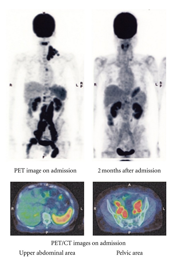
Imaging test results: the coronal PET image (upper left) shows wide spread pathological uptake of radiotracer (FDG) in cervical, supraclavicular, mediastinal, para-aortic, peri-iliac and inguinal lymph nodes and spleen. A second PET/CT scan performed 2 months after admission (upper right) shows complete resolution of the abnormal FDG activity. Fused PET/CT image on admission (lower left) shows moderate FDG uptake in the spleen.
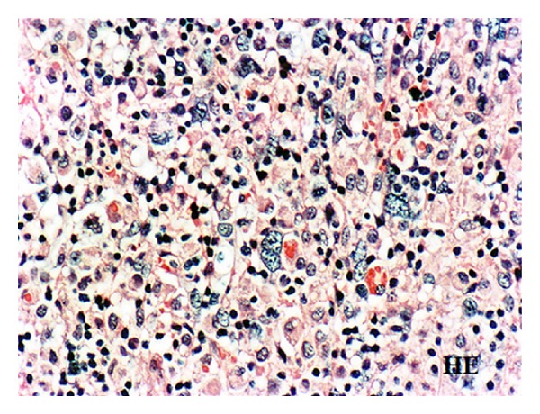
Histology of the cervical lymph node biopsy showing the infiltration of giant cells reminiscent of Reed-Sternberg cells and atypical large round cells with remarkable nucleoli in the background of lymphocytes, original magnification ×300. H&E stain.
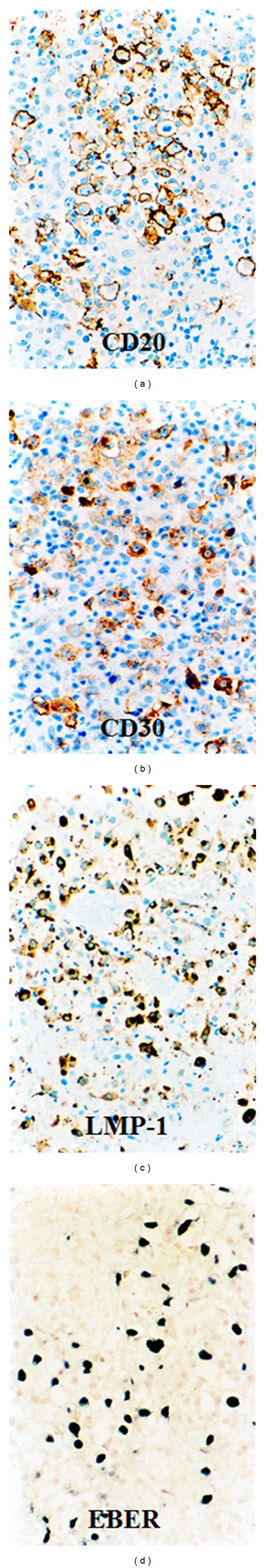
Immunohistochemical stainings for CD20, CD30 and LMP-1 and EBER staining (in situ hybridization). Tumor cells are positive for CD20, CD30, LMP-1 and EBER, original magnification ×300.
Similar articles
-
Methotrexate-related Epstein-Barr Virus (EBV)-associated lymphoproliferative disorder--so-called "Hodgkin-like lesion"--of the oral cavity in a patient with rheumatoid arthritis.Head Neck Pathol. 2010 Dec;4(4):305-11. doi: 10.1007/s12105-010-0202-6. Epub 2010 Jul 31. Head Neck Pathol. 2010. PMID: 20676828 Free PMC article.
-
Remission of lymphoma after withdrawal of methotrexate in rheumatoid arthritis: relationship with type of latent Epstein-Barr virus infection.Am J Hematol. 2007 Dec;82(12):1106-9. doi: 10.1002/ajh.21003. Am J Hematol. 2007. PMID: 17654684
-
Successful Treatment of Intracranial Methotrexate-associated Lymphoproliferative Disorder without Epstein-Barr Virus Infection Using Rituximab, Methotrexate, Procarbazine, and Vincristine: A Case Report.NMC Case Rep J. 2022 Jul 27;9:237-242. doi: 10.2176/jns-nmc.2022-0091. eCollection 2022. NMC Case Rep J. 2022. PMID: 36061907 Free PMC article.
-
Spontaneous regression of lymphoproliferative disorders in patients treated with methotrexate for rheumatoid arthritis and other rheumatic diseases.J Clin Oncol. 1996 Jun;14(6):1943-9. doi: 10.1200/JCO.1996.14.6.1943. J Clin Oncol. 1996. PMID: 8656264 Review.
-
Methotrexate-associated lymphoproliferative disorders of the tongue developing in patients with rheumatoid arthritis: a report of 2 cases and a review.Oral Surg Oral Med Oral Pathol Oral Radiol. 2015 Jan;119(1):e1-5. doi: 10.1016/j.oooo.2014.04.002. Epub 2014 Apr 18. Oral Surg Oral Med Oral Pathol Oral Radiol. 2015. PMID: 24927637 Review.
Cited by
-
Possible mechanisms of action of clarithromycin and its clinical application as a repurposing drug for treating multiple myeloma.Ecancermedicalscience. 2020 Aug 18;14:1088. doi: 10.3332/ecancer.2020.1088. eCollection 2020. Ecancermedicalscience. 2020. PMID: 33014130 Free PMC article. Review.
-
Treatment of advanced stage methotrexate-associated lymphoproliferative disorders (MTX-LPDs) with methotrexate discontinuation.BMJ Case Rep. 2018 Dec 13;11(1):e226545. doi: 10.1136/bcr-2018-226545. BMJ Case Rep. 2018. PMID: 30567235 Free PMC article.
-
Successful treatment in a case of Propionibacterium acnes-associated sarcoidosis with clarithromycin administration: a case report.J Med Case Rep. 2014 Jan 15;8:15. doi: 10.1186/1752-1947-8-15. J Med Case Rep. 2014. PMID: 24428939 Free PMC article.
-
Does it take three to tango? An unsuspected multimorbidity of CD8+ T cell lymphoproliferative disorder, malaria, and EBV infection.Malar J. 2018 Oct 5;17(1):349. doi: 10.1186/s12936-018-2497-9. Malar J. 2018. PMID: 30290813 Free PMC article.
-
Clinical management for other iatrogenic immunodeficiency-associated lymphoproliferative disorders.J Clin Exp Hematop. 2019;59(2):72-92. doi: 10.3960/jslrt.19007. J Clin Exp Hematop. 2019. PMID: 31257348 Free PMC article. Review.
References
-
- Hakulinen T, Isomaki H, Knekt P. Rheumatoid arthritis and cancer studies based on linking nationwide registries in Finland. The American Journal of Medicine. 1985;78(1):29–32. - PubMed
-
- Gridley G, McLaughlin JK, Ekbom A, et al. Incidence of cancer among patients with rheumatoid arthritis. Journal of the National Cancer Institute. 1993;85(4):307–311. - PubMed
-
- Mellemkjær L, Linet MS, Gridley G, Frisch M, Møller H, Olsen JH. Rheumatoid arthritis and cancer risk. European Journal of Cancer. A. 1996;32(10):1753–1757. - PubMed
-
- Ellman MH, Hurwitz H, Thomas C, Kozloff M. Lymphoma developing in a patient with rheumatoid arthritis taking low dose weekly methotrexate. The Journal of Rheumatology. 1991;18(11):1741–1743. - PubMed
-
- Moder KG, Tefferi A, Cohen MD, Menke DM, Luthra HS. Hematologic malignancies and the use of methotrexate in rheumatoid arthritis: a retrospective study. The American Journal of Medicine. 1995;99(3):276–281. - PubMed
LinkOut - more resources
Full Text Sources
