Comparative Study
. 2013 Jan;3(1):41-63.
doi: 10.1534/g3.112.004044. Epub 2013 Jan 1.

Comparative genomics of a plant-pathogenic fungus, Pyrenophora tritici-repentis, reveals transduplication and the impact of repeat elements on pathogenicity and population divergence

Affiliations
Comparative Study

Comparative genomics of a plant-pathogenic fungus, Pyrenophora tritici-repentis, reveals transduplication and the impact of repeat elements on pathogenicity and population divergence

Viola A Manning et al. G3 (Bethesda). 2013 Jan.

Abstract

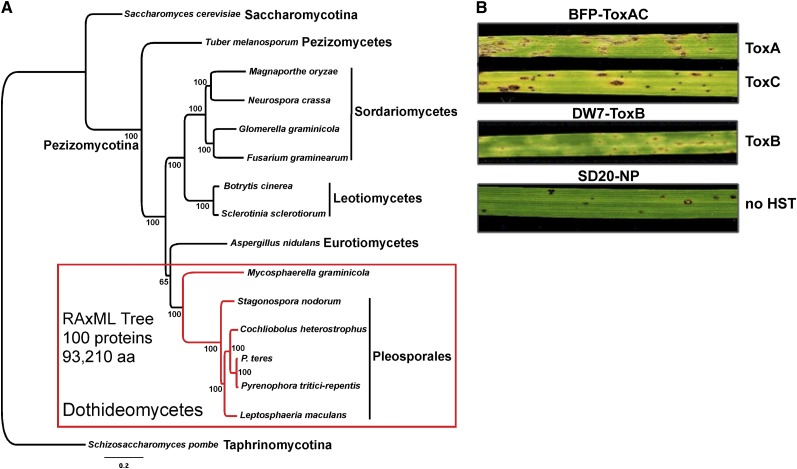
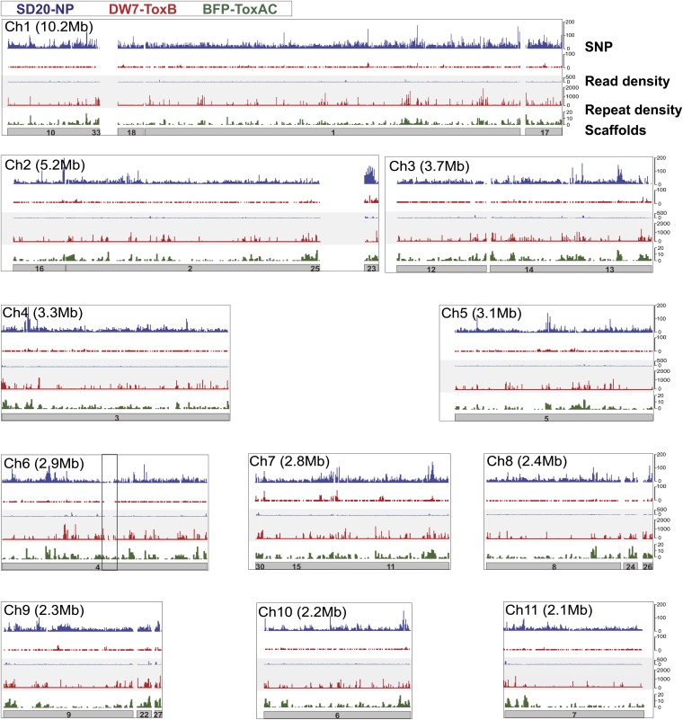
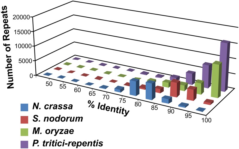
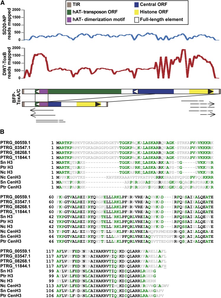
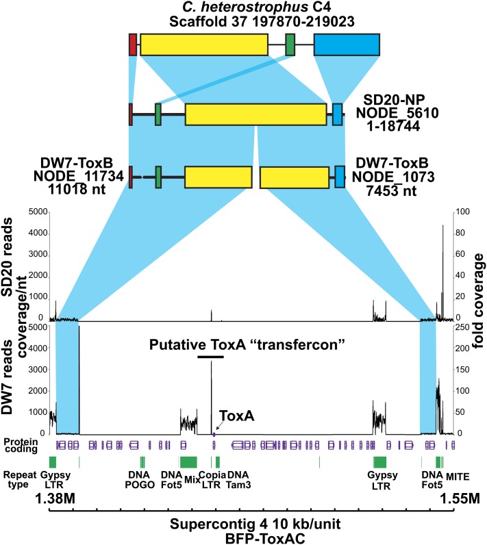
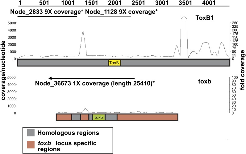
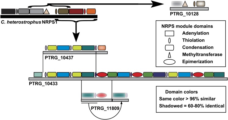
Pyrenophora tritici-repentis is a necrotrophic fungus causal to the disease tan spot of wheat, whose contribution to crop loss has increased significantly during the last few decades. Pathogenicity by this fungus is attributed to the production of host-selective toxins (HST), which are recognized by their host in a genotype-specific manner. To better understand the mechanisms that have led to the increase in disease incidence related to this pathogen, we sequenced the genomes of three P. tritici-repentis isolates. A pathogenic isolate that produces two known HSTs was used to assemble a reference nuclear genome of approximately 40 Mb composed of 11 chromosomes that encode 12,141 predicted genes. Comparison of the reference genome with those of a pathogenic isolate that produces a third HST, and a nonpathogenic isolate, showed the nonpathogen genome to be more diverged than those of the two pathogens. Examination of gene-coding regions has provided candidate pathogen-specific proteins and revealed gene families that may play a role in a necrotrophic lifestyle. Analysis of transposable elements suggests that their presence in the genome of pathogenic isolates contributes to the creation of novel genes, effector diversification, possible horizontal gene transfer events, identified copy number variation, and the first example of transduplication by DNA transposable elements in fungi. Overall, comparative analysis of these genomes provides evidence that pathogenicity in this species arose through an influx of transposable elements, which created a genetically flexible landscape that can easily respond to environmental changes.

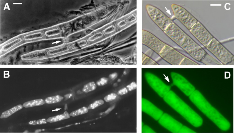
Keywords: ToxA; ToxB; anastomosis; copy number variation; histone H3 transduplication; wheat (Triticum aestivum).


Figures

Figure 1
Figure 1
Phylogeny of Pyrenophora tritici-repentis and symptoms induced by the three sequenced isolates. RAxML maximum likelihood phylogenetic tree (best tree -nL1505566.555282) inferred from a 100-protein superalignment comprising 93,210 amino acid positions (A). Subphyla (-mycotina) and classes (-mycetes) of the phylum Ascomycota are shown and numbers near nodes are bootstrap partitions. (B) ToxA and ToxC symptoms induced by BFP-ToxAC (on Glenlea and 6B365, respectively; top 2 leaves), ToxB symptoms induced by DW7-ToxB (on 6B662; middle leaf), and the resistant reaction produced by the nonpathogenic SD20-NP (on Auburn; bottom leaf).
Figure 2
Figure 2
Mapping of sequence reads of resequenced isolates relative to the reference genome of Pyrenophora tritici-repentis. Schematic represents the reference genome scaffolds (gray bars with supercontig numbers) mapped to each chromosome as defined by the reference optical map. The border box reflects chromosome size as indicated next to the chromosome number and white spaces between the scaffolds represent gaps in the sequence assemblies. Repeat density (green) in the reference isolate, and read (gray central panel) and SNP (top panel) densities of the Illumina-sequenced isolates (DW7-ToxB-red, SD20-NP-blue), were mapped per 10 kb of the high-quality genome assembly based on the reference isolate (BFP-ToxAC) of P. tritici-repentis.
Figure 3
Figure 3
Repeat identity in the genome sequence of Pyrenophora tritici-repentis. The graph represents a comparison of repeat similarities between Neurospora crassa, Stagonospora nodorum, Magnaporthe oryzae, and Pyrenophora tritici-repentis. An identity was assigned to each repetitive sequence based on its similarity to other repeats in the genome (naturally grouped into one repeat family). The number shown on the y-axis is a collection of all repeats identified in the genome sequences (minimal 400 bp, 50% identity) and binned into groups according to the sequence identity present in that repeat family.
Figure 4
Figure 4
Conidia of Pyrenophora tritici-repentis connected by anastomosis bridges. (A) Phase-contrast microscopy of conidial anastomosis in Ptr. (B) The same conidia shown in A stained with DAPI and visualized under an ultraviolet filter to image nuclei; Note the loss of nuclei from the upper cell. (C) Nomarski optics (D) and fluorescence microscopy of anastomosed conidia of Ptr expressing green fluorescent protein. Arrows indicate the anastomosis bridges. Scale bars = approximately 5 µm.
Figure 5
Figure 5
Transduplication of a histone H3-like protein family in the genome of Pyrenophora tritici-repentis. (A) Two representatives of a transduplicating family of DNA transposable elements in the BFP-ToxAC reference genome are shown with the characteristic motifs of the elements indicated (brown box = terminal inverted repeats (TIR); green box = hAT transposon coding regions; purple box = hAT dimerization motif; blue box = central open reading frame (ORF) coding region; yellow box = histone H3 coding region; black boundary box = full-length element). The large, likely autonomous element is approximately 5.6 kb in length. Black lines between elements illustrate a pathogen-specific recombination event that results in a smaller element of approximately 2.3 kb. The graphs show coverage of reads obtained from the pathogenic DW7-ToxB and nonpathogenic SD20-NP isolates. Arrows and lines below the element representation indicate alignments of ESTs detected in various libraries (arrows = poly(A) tails, light gray = introns). (B) Alignment of novel histone H3-like (H3L) proteins identified in Ptr with bona fide histone H3 variants from Ptr, S. nodorum, and N. crassa. Four variants of full-length H3L exist in Ptr, all are different by a single aa change (underlined; also see Table S9). Two changes in H3L alleles result in frameshift mutations, yielding 5′ and 3′ truncated versions of H3L. Most (21) copies are identical to PTRG_00559.1 in amino acid and DNA sequence. Completely conserved residues are shown in black, similar residues in green and variable residues in gray.
Figure 6
Figure 6
Absence of the 145-kb ToxA-containing region in the pathogenic DW7-ToxB and non-pathogenic SD20-NP isolates of Pyrenophora tritici-repentis. Schematic includes the protein-coding regions (purple) and repeats (green) present within the 170-kb ToxA-containing genomic region in the BFP-ToxAC reference genome. The graphs show coverage of reads obtained from the pathogenic DW7-ToxB and nonpathogenic SD20-NP isolates. Coverage depth per nucleotide (nt) is indicated on the left and fold coverage is indicated on the right. Blue shading represents synteny between genomic scaffolds of Cochliobolus heterostrophus isolate C4, SD20-NP, DW7-ToxB, and flanking regions of the 170-kb ToxA-containing region of the reference. Similarly colored bars within the scaffolds indicate colinear blocks.
Figure 7
Figure 7
Read mapping to and de novo assembly of ToxB- and toxb-containing loci in the genome of Pyrenophora tritici-repentis. Schematic of Illumina sequence reads (line graph) of isolate DW7-ToxB mapped to the ToxB1 locus (top: ToxB1 locus; accession number: AY425480.1) and of SD20-NP mapped to the toxb locus (bottom: toxb locus; accession number: AY083456.2). Coverage depth per nt is indicated on the left and fold coverage is indicated on the right. Straight lines above the graphs depict the contigs present in the de novo assemblies of the Illumina-sequenced isolates. The arrow on the contig above the toxb locus shows how that contig extends beyond the locus.
Figure 8
Figure 8
NRPSs diversification in the genome of Pyrenophora tritici-repentis. Schematic shows the modular architecture of Cochliobolus heterostrophus NRPS1 (black solid bar and associated modules) and related P. tritici-repentis PtNRPSs (gray solid bars and associated modules). Brackets with straight arrows define modules in C. heterostrophus NRPS1 that have homologous and/or related regions present in PtNRPSs. The module domains (adenylation, thiolation, condensation, methyltransferase, and epimerization), and the solid bars that represent the entire protein are drawn to scale. Thin lines between gray bars extend to the region(s) of identity between proteins and the curved arrow depicts a recent duplication event.
Figure 9
Figure 9
Predicted secreted proteins in the genome sequence of Pyrenophora tritici-repentis. (A) Distribution of secreted proteins in the reference Ptr isolate (BFP-ToxAC) and the Illumina-sequenced pathogenic (DW7-ToxB) and nonpathogenic (SD20-NP) isolates. (B) PCR screen of pathogenic and nonpathogenic isolates for the presence of the gene and transcript for PTRG_11888, a predicted putative secreted protein. Pathogenic isolates: BFP-ToxAC; ASC1; 86-124; D308; DW2; SO3-P; Non-pathogenic isolates: SD20-NP; 90-2; 98-31-2 (see also Table S1).

References

    1. Abascal F., Zardoya R., Posada D., 2005. ProtTest: selection of best-fit models of protein evolution. Bioinformatics 21: 2104–2105 - PubMed
    1. Aboukhaddour R., Cloutier S., Ballance G. M., Lamari L., 2009. Genome characterization of Pyrenophora tritici-repentis isolates reveals high plasticity and independent chromosomal location of ToxA and ToxB. Mol. Plant Pathol. 10: 201–212 - PMC - PubMed
    1. Aboukhaddour R., Cloutier S., Lamari L., Strelkov S. E., 2011. Simple sequence repeats and diversity of globally distributed populations of Pyrenophora tritici-repentis. Can. J. Plant Pathol. 33: 389–399
    1. Aboukhaddour R., Kim Y. M., Strelkov S. E., 2012. RNA-mediated gene silencing of ToxB in Pyrenophora tritici-repentis. Mol. Plant Pathol. 13: 318–326 - PMC - PubMed
    1. Akagi Y., Akamatsu H., Otani H., Kodama M., 2009. Horizontal chromosome transfer, a mechanism for the evolution and differentiation of a plant-pathogenic fungus. Eukaryot. Cell 8: 1732–1738 - PMC - PubMed

Publication types

MeSH terms