

Role of FDG-PET scans in staging, response assessment, and follow-up care for non-small cell lung cancer
- PMID: 23316478
- PMCID: PMC3539654
- DOI: 10.3389/fonc.2012.00208
Role of FDG-PET scans in staging, response assessment, and follow-up care for non-small cell lung cancer
Abstract
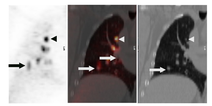
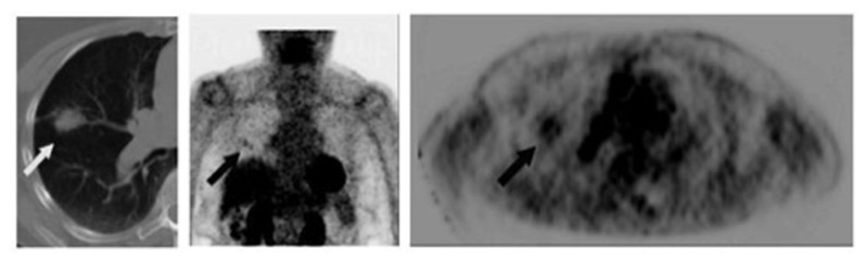
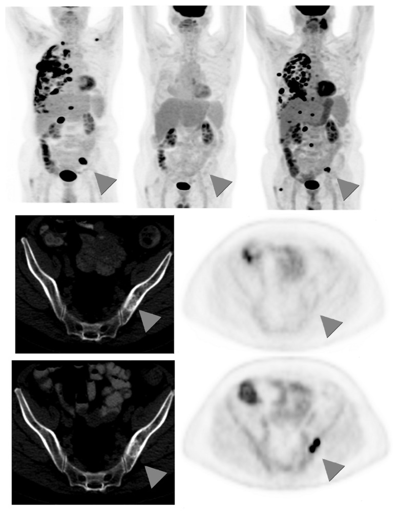
The integral role of positron-emission tomography (PET) using the glucose analog tracer fluorine-18 fluorodeoxyglucose (FDG) in the staging of non-small cell lung cancer (NSCLC) is well established. Evidence is emerging for the role of PET in response assessment to neoadjuvant therapy, combined-modality therapy, and early detection of recurrence. Here, we review the current literature on these aspects of PET in the management of NSCLC. FDG-PET, particularly integrated (18)F-FDG-PET/CT, scans have become a standard test in the staging of local tumor extent, mediastinal lymph node involvement, and distant metastatic disease in NSCLC. (18)F-FDG-PET sensitivity is generally superior to computed tomography (CT) scans alone. Local tumor extent and T stage can be more accurately determined with FDG-PET in certain cases, especially in areas of post-obstructive atelectasis or low CT density variation. FDG-PET sensitivity is decreased in tumors <1 cm, at least in part due to respiratory motion. False-negative results can occur in areas of low tumor burden, e.g., small lymph nodes or ground-glass opacities. (18)F-FDG-PET-CT nodal staging is more accurate than CT alone, as hilar and mediastinal involvement is often detected first on (18)F-FDG-PET scan when CT criteria for malignant involvement are not met. (18)F-FDG-PET scans have widely replaced bone scintography for assessing distant metastases, except for the brain, which still warrants dedicated brain imaging. (18)F-FDG uptake has also been shown to vary between histologies, with adenocarcinomas generally being less FDG avid than squamous cell carcinomas. (18)F-FDG-PET scans are useful to detect recurrences, but are currently not recommended for routine follow-up. Typically, patients are followed with chest CT scans every 3-6 months, using (18)F-FDG-PET to evaluate equivocal CT findings. As high (18)F-FDG uptake can occur in infectious, inflammatory, and other non-neoplastic conditions, (18)F-FDG-PET-positive findings require pathological confirmation in most cases. There is increased interest in the prognostic and predictive role of FDG-PET scans. Studies show that absence of metabolic response to neoadjuvant therapy correlates with poor pathologic response, and a favorable (18)F-FDG-PET response appears to be associated with improved survival. Further work is underway to identify subsets of patients that might benefit individualized management based on FDG-PET.
Keywords: PET; follow-up; non-small cell lung cancer; response assessment; staging.
Figures
Similar articles
-
More advantages in detecting bone and soft tissue metastases from prostate cancer using 18F-PSMA PET/CT.Hell J Nucl Med. 2019 Jan-Apr;22(1):6-9. doi: 10.1967/s002449910952. Epub 2019 Mar 7. Hell J Nucl Med. 2019. PMID: 30843003
-
Mediastinal lymph node staging by FDG-PET in patients with non-small cell lung cancer: analysis of false-positive FDG-PET findings.Respiration. 2003 Sep-Oct;70(5):500-6. doi: 10.1159/000074207. Respiration. 2003. PMID: 14665776
-
Combined endobronchial and esophageal endosonography for the diagnosis and staging of lung cancer: European Society of Gastrointestinal Endoscopy (ESGE) Guideline, in cooperation with the European Respiratory Society (ERS) and the European Society of Thoracic Surgeons (ESTS).Endoscopy. 2015 Jun;47(6):545-59. doi: 10.1055/s-0034-1392040. Epub 2015 Jun 1. Endoscopy. 2015. PMID: 26030890
-
Overview of the clinical effectiveness of positron emission tomography imaging in selected cancers.Health Technol Assess. 2007 Oct;11(44):iii-iv, xi-267. doi: 10.3310/hta11440. Health Technol Assess. 2007. PMID: 17999839 Review.
-
Role of 18F-FDG PET/CT in establishing new clinical and therapeutic modalities in lung cancer. A short review.Rev Esp Med Nucl Imagen Mol (Engl Ed). 2019 Jul-Aug;38(4):229-233. doi: 10.1016/j.remn.2019.02.003. Epub 2019 Jun 13. Rev Esp Med Nucl Imagen Mol (Engl Ed). 2019. PMID: 31202725 Review. English, Spanish.
Cited by
-
Safety and Efficacy of Lattice Radiotherapy in Voluminous Non-small Cell Lung Cancer.Cureus. 2019 Mar 18;11(3):e4263. doi: 10.7759/cureus.4263. Cureus. 2019. PMID: 31139522 Free PMC article.
-
The role of positron emission tomography in the diagnosis, staging and response assessment of non-small cell lung cancer.Ann Transl Med. 2018 Mar;6(5):95. doi: 10.21037/atm.2018.01.25. Ann Transl Med. 2018. PMID: 29666818 Free PMC article. Review.
-
Prognostic Value of Metabolic Tumor Parameters in Pretreatment 18F-Fluorodeoxyglucose Positron Emission Tomography-Computed Tomography Scan in Advanced Non-Small Cell Lung Cancer.Indian J Nucl Med. 2021 Apr-Jun;36(2):107-113. doi: 10.4103/ijnm.IJNM_170_20. Epub 2021 Jun 21. Indian J Nucl Med. 2021. PMID: 34385779 Free PMC article.
-
Predictive risk score for isolated brain metastasis in non-small cell lung cancer.J Thorac Dis. 2024 Jun 30;16(6):3794-3804. doi: 10.21037/jtd-23-1668. Epub 2024 Jun 19. J Thorac Dis. 2024. PMID: 38983167 Free PMC article.
-
Positron emission tomography imaging of lung cancer: An overview of alternative positron emission tomography tracers beyond F18 fluorodeoxyglucose.Front Med (Lausanne). 2022 Oct 5;9:945602. doi: 10.3389/fmed.2022.945602. eCollection 2022. Front Med (Lausanne). 2022. PMID: 36275809 Free PMC article. Review.
References
-
- Al-Sarraf N., Gately K., Lucey J., Wilson L., Mcgovern E., Young V. (2008). Lymph node staging by means of positron emission tomography is less accurate in non-small cell lung cancer patients with enlarged lymph nodes: analysis of 1,145 lymph nodes. Lung Cancer 60 62–68 - PubMed
-
- Aquino S. L., Halpern E. F., Kuester L. B., Fischman A. J. (2007). FDG-PET and CT features of non-small cell lung cancer based on tumor type. Int. J. Mol. Med. 19 495–499 - PubMed
-
- Berghmans T., Dusart M., Paesmans M., Hossein-Foucher C., Buvat I., Castaigne C., et al. (2008). Primary tumor standardized uptake value (SUVmax) measured on fluorodeoxyglucose positron emission tomography (FDG-PET) is of prognostic value for survival in non-small cell lung cancer (NSCLC): a systematic review and meta-analysis (MA) by the European Lung Cancer Working Party for the IASLC Lung Cancer Staging Project. J. Thorac. Oncol. 3 6–12 - PubMed
-
- Burdick M. J., Stephans K. L., Reddy C. A., Djemil T., Srinivas S. M., Videtic G. M. (2010). Maximum standardized uptake value from staging FDG-PET/CT does not predict treatment outcome for early-stage non-small-cell lung cancer treated with stereotactic body radiotherapy. Int. J. Radiat. Oncol. Biol. Phys. 78 1033–1039 - PubMed
-
- Bury T., Barreto A., Daenen F., Barthelemy N., Ghaye B., Rigo P. (1998). Fluorine-18 deoxyglucose positron emission tomography for the detection of bone metastases in patients with non-small cell lung cancer. Eur. J. Nucl. Med. 25 1244–1247 - PubMed
Grants and funding
LinkOut - more resources
Full Text Sources
Other Literature Sources
Research Materials
Miscellaneous