. 2013 Jan 14:8:2.
doi: 10.1186/1750-1326-8-2.

Neuron loss in the 5XFAD mouse model of Alzheimer's disease correlates with intraneuronal Aβ42 accumulation and Caspase-3 activation

Affiliations

Neuron loss in the 5XFAD mouse model of Alzheimer's disease correlates with intraneuronal Aβ42 accumulation and Caspase-3 activation

William A Eimer et al. Mol Neurodegener. .

Abstract

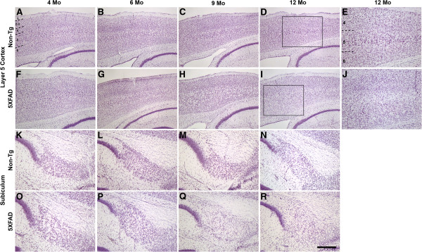
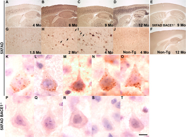
Background: Although the mechanism of neuron loss in Alzheimer's disease (AD) is enigmatic, it is associated with cerebral accumulation of Aβ42. The 5XFAD mouse model of amyloid deposition expresses five familial AD (FAD) mutations that are additive in driving Aβ42 overproduction. 5XFAD mice exhibit intraneuronal Aβ42 accumulation at 1.5 months, amyloid deposition at 2 months, and memory deficits by 4 months of age.

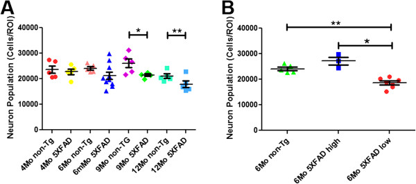
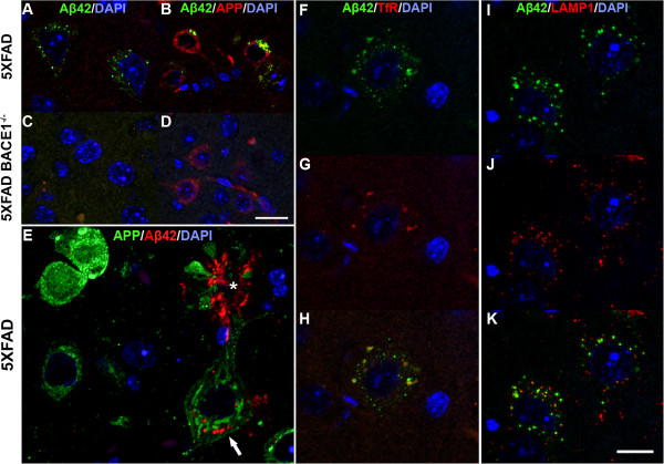
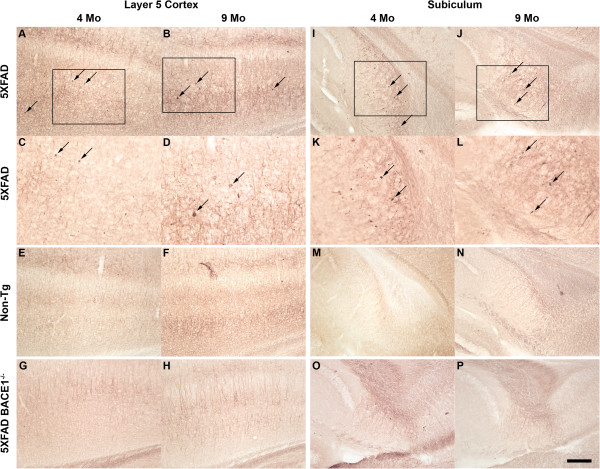
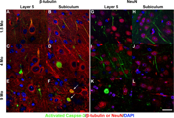
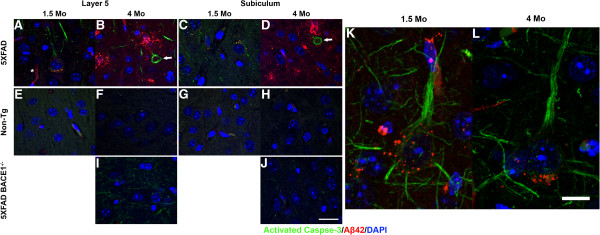
Results: Here, we demonstrate by unbiased stereology that statistically significant neuron loss occurs by 9 months of age in 5XFAD mice. We validated two Aβ42-selective antibodies by immunostaining 5XFAD; BACE1-/- bigenic brain sections and then used these antibodies to show that intraneuronal Aβ42 and amyloid deposition develop in the same regions where neuron loss is observed in 5XFAD brain. In 5XFAD neuronal soma, intraneuronal Aβ42 accumulates in puncta that co-label for Transferrin receptor and LAMP-1, indicating endosomal and lysosomal localization, respectively. In addition, in young 5XFAD brains, we observed activated Caspase-3 in the soma and proximal dendrites of intraneuronal Aβ42-labeled neurons. In older 5XFAD brains, we found activated Caspase-3-positive punctate accumulations that co-localize with the neuronal marker class III β-tubulin, suggesting neuron loss by apoptosis.

Conclusions: Together, our results indicate a temporal sequence of intraneuronal Aβ42 accumulation, Caspase-3 activation, and neuron loss that implies a potential apoptotic mechanism of neuron death in the 5XFAD mouse.


Figures

Figure 1
Figure 1
5XFAD mice exhibit neuron loss in Layer 5 cortex and subiculum. Parasagittal brain sections from representative 4- to 12-month-old non-transgenic littermate (A-E, K-N) and 5XFAD (F-J, O-R) female mice were stained with cresyl violet and micrographed to image Layer 5 cortex (A-J) and subiculum (K-R). Numbers indicate cortical layers and dashed lines identify borders between layers. Boxes in D and I outline the areas of increased magnification shown in E and J, respectively. The 5XFAD mouse exhibits visible loss of large pyramidal neurons at 9 months of age as evident by a decrease in stained neurons (H, Q, I, R, J). Increased magnification (J) shows a clear loss of large pyramidal neurons in Layer 5 of the 5XFAD mouse at 12 months compared to the non-transgenic (E). Scale bar in R = 280 μm for A-D, F-I; 110 μm for E, J; 80 μm for K-R.
Figure 2
Figure 2
Unbiased stereological analysis of 5XFAD mice reveals significant neuron loss in cortical Layer 5 at 9 and 12 months of age. Brains from age-matched non-transgenic littermate (non-Tg) and 5XFAD female mice were sectioned at 30 μm and stained with cresyl violet and imaged by light microscopy. Large pyramidal neurons were then counted in Layer 5 cortex using an optical fractionator. (A) Large pyramidal neuron numbers in non-transgenic and 5XFAD Layer 5 cortex from mice aged 4–12 months. At 9 and 12 months there is a statistically significant decrease in the number of large pyramidal neurons in 5XFAD compared to non-transgenic mice (* p<0.05, ** p<0.01; n = 5 mice per group). 5XFAD mice exhibited a progressive decrease in Layer 5 large pyramidal neuron number, which at 12 months represented a 24.8% loss compared to non-transgenic mice. (B) 6 month 5XFAD mice are separable into two subgroups: those with normal neuron counts (6Mo 5XFAD high) and those with low neuron counts indicating neuron loss (6Mo 5XFAD low). The 5XFAD low count group showed a statistically significant decrease in neuron number compared to age matched non-transgenic and 5XFAD normal count groups (* p<0.05, ** p<0.01, n = 5 non-Tg, n = 10 net 6 month 5XFAD).
Figure 3
Figure 3
5XFAD mice exhibit intraneuronal Aβ42 prior to amyloid deposition and neuron loss. Parasagittal brain sections from 1.5 (G), 2 (H), 4 (A, I-T), 6 (B), 9 (C, E), and 12 (D, F) month mice were incubated with anti-Aβ42 C-terminal neo-epitope antibody, visualized by DAB staining, and micrographed. 5XFAD mice display progressive Aβ42-positive plaque deposition with age (A-D). In 5XFAD Layer 5 cortex, intraneuronal Aβ42 (arrows) is first observed at 1.5 months (G) and amyloid plaques at 2 months (H) of age. At high magnification, intraneuronal Aβ42 staining is confined within numerous small spherical puncta dispersed throughout the cytoplasm of the soma of large pyramidal neurons (K-O). The appearance of these Aβ42-positive puncta suggests membrane-bound intracellular compartments such as endosomes or lysosomes. Importantly, the absence of Aβ42 staining in 5XFAD; BACE1−/− neurons (E, P-T) demonstrates the selectivity of the antibody for Aβ42, and indicates that the antibody dos not cross-react with endogenous or transgenic APP under the conditions used. Aβ42 staining is also lacking in non-transgenic littermate (Non-Tg) sections (F, J). Scale bar = 400 μm (A-F), 40 μm (G-J), 9 μm (K-T).
Figure 4
Figure 4
Intraneuronal Aβ42 is partially localized within both endosomes and lysosomes in 5XFAD neurons. Parasagittal brain sections of 5XFAD (A, B, E-K) and 5XFAD; BACE1−/− (C, D) brains were co-stained with anti-Aβ42-selective antibody (A-K) and anti-APP (6E10: B, D; Karen [29]: E) or anti-Transferrin receptor (TfR) (F-H) or anti-LAMP1 (I-K) antibody, and DAPI for nuclei (A-K), and imaged by confocal microscopy. All mice were 4 months old, except mice used for LAMP1 immunostaining were 2 months (I-K). Label colors indicate fluorescence color. Note that APP staining appears widely distributed in the soma and cell surface (B, D, E), while Aβ42 labeling is confined to intraneuronal puncta in 5XFAD mice (A, B, E, F, I). Asterisk in E identifies Aβ42-positive plaque (red) above large pyramidal neuron that exhibits intraneuronal Aβ42 puncta (arrow). lntraneuronal Aβ42 signal is absent in 5XFAD; BACE1−/− neurons (C, D), again demonstrating the Aβ42-selectivity of the antibody. In 5XFAD neurons, intraneuronal Aβ42 co-localizes with Transferrin receptor in early endosomes (F-H) and with LAMP1 in late endosomes and early lysosomes (I-K), at least in part. Scale bar in D = 12μm (A, C); = 20 μm (B, D). Scale bar in K = 12 μm (E); = 20 μm (F-K).
Figure 5
Figure 5
5XFAD mice exhibit Caspase-3 activation in Layer 5 cortex and subiculum. Parasagittal brain sections from representative 4 and 9 month old 5XFAD (A-D, I-L), non-transgenic littermate (Non-Tg; E, F, M, N), and 5XFAD; BACE1−/− (G, H, O, P) mice were incubated with an antibody against activated Caspase-3, visualized by DAB staining, and micrographed in Layer 5 cortex (A-H) and subiculum (I-P). The levels of activated Caspase-3 immunolabeling are significantly higher in the brains of both ages of 5XFAD mice compared to those of 5XFAD; BACE1−/− or non-transgenic mice. In addition, punctate aggregates of activated Caspase-3 are distributed throughout 5XFAD sections (black arrows, A-D, I-L), but are absent in 5XFAD; BACE1−/− or Non-Tg sections (E-H, M-P). Boxes in A, B, I, and J are enlarged in C, D, K, and L, respectively. Scale bar A, B, E-H, I, J, M-P = 80 μm; C, D, K, L = 40 μm.
Figure 6
Figure 6
5XFAD mice have elevated levels of activated Caspase-3 in neurons of Layer 5 cortex and subiculum. Parasagittal brain sections from 1.5, 4, and 9 month old 5XFAD mice were co-incubated with antibodies against activated Caspase-3 (green) and neuron-specific class III β-tubulin (red; A-F) or NeuN (red; G-L) and imaged by confocal microscopy. Sections were co-stained with DAPI to detect nuclei (blue). Co-immunolabeling with neuronal markers β-tubulin and NeuN demonstrate Caspase-3 activation in 5XFAD neurons. Note the presence of activated Caspase-3 labeling in proximal dendrites of large pyramidal neurons of Layer 5 cortex and subiculum at 1.5 and 4 months (A-D, G-J), The number of Caspase-3-positive proximal dendrites is reduced in 9 month old compared to younger 5XFAD mice, although Caspase-3 aggregates are more prevalent at older ages suggesting a progressive process (spherical green structures in E, F, K, L). Activated Caspase-3-positive aggregates become apparent at 4 months in Layer 5 cortex and later in subiculum, and likely represent neuronal apoptotic bodies. Arrows in F point to Caspase-3-positive aggregates that co-label with neuron-specific class III β-tubulin. Scale bar = 20 μm.
Figure 7
Figure 7
5XFAD mice display Caspase-3 activation in neurons with intraneuronal Aβ42. Parasagittal brain sections from 1.5 (A, E, C, G, K) and 4 month (B, D, F, H-J, L) 5XFAD (A-D, K, L), non-transgenic littermate (Non-Tg; E-H), and 5XFAD; BACE1−/− (I, J) mice were co-incubated with antibodies against activated Caspase-3 (green) and Aβ42 C-terminal neo-epitope (red) and imaged by confocal microscopy. Sections were co-stained with DAPI to detect nuclei (blue). Activated Caspase-3 is present in 5XFAD large pyramidal neurons of Layer 5 cortex (A, B, K, L) and subiculum (C, D) that also display intraneuronal Aβ42. At high magnification, activated Caspase-3 is distributed throughout in the soma and proximal dendrites of Aβ42-positive neurons (K, L). Neither activated Caspase-3 nor intraneuronal Aβ42 are present in neurons of Non-Tg (E-H) or 5XFAD; BACE1−/− (I, J) mice. Note presence of amyloid plaques (red) at 4 months in 5XFAD brain (B, D). Activated Caspase-3-positive ring-like structures (arrows, B, D) may represent neurons in the process of degeneration. Asterisk in A identifies background capillary staining. Scale bar A-J = 20 μm; K, L = 10 μm.

References

    1. Bertram L, Lill CM, Tanzi RE. The genetics of Alzheimer disease: back to the future. Neuron. 2010;68:270–281. doi: 10.1016/j.neuron.2010.10.013. - DOI - PubMed
    1. Sisodia SS. Alzheimer’s disease: perspectives for the new millennium. J Clin Invest. 1999;104:1169–1170. doi: 10.1172/JCI8508. - DOI - PMC - PubMed
    1. Hyslop PA, Bender MH. Methods for sample preparation for direct immunoassay measurement of analytes in tissue homogenates: ELISA assay of amyloid beta-peptides. Curr Protoc Neurosci. 2002;Chapter 7:Unit 7 20. - PubMed
    1. Selkoe D, Mandelkow E, Holtzman D. Deciphering Alzheimer disease. Cold Spring Harb Perspect Med. 2012;2:a011460. - PMC - PubMed
    1. Holtzman DM, Morris JC, Goate AM. Alzheimer’s disease: the challenge of the second century. Sci Transl Med. 2011;3:77sr71. - PMC - PubMed

Publication types