. 2013 Feb 14;56(3):867-78.
doi: 10.1021/jm301338q. Epub 2013 Jan 28.

Sulfated pentagalloylglucoside is a potent, allosteric, and selective inhibitor of factor XIa

Affiliations

Sulfated pentagalloylglucoside is a potent, allosteric, and selective inhibitor of factor XIa

Rami A Al-Horani et al. J Med Chem. .

Abstract

Inhibition of factor XIa (FXIa) is a novel paradigm for developing anticoagulants without major bleeding consequences. We present the discovery of sulfated pentagalloylglucoside (6) as a highly selective inhibitor of human FXIa. Biochemical screening of a focused library led to the identification of 6, a sulfated aromatic mimetic of heparin. Inhibitor 6 displayed a potency of 551 nM against FXIa, which was at least 200-fold more selective than other relevant enzymes. It also prevented activation of factor IX and prolonged human plasma and whole blood clotting. Inhibitor 6 reduced V(MAX) of FXIa hydrolysis of chromogenic substrate without affecting the K(M), suggesting an allosteric mechanism. Competitive studies showed that 6 bound in the heparin-binding site of FXIa. No allosteric small molecule has been discovered to date that exhibits equivalent potency against FXIa. Inhibitor 6 is expected to open up a major route to allosteric FXIa anticoagulants with clinical relevance.


Figures
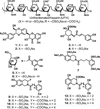
Figure 1
Figure 1. Structures of heparins (A) and the polyphenolic as well as the sulfated molecules were screened against factor XIa (B)
Shown in A are heparins including unfractionated heparin (UFH) (Mwt ~ 15,000 Da) as well as LMWHs (Mwt ~ 4000 Da), both of which are highly heterogeneous and polydisperse mixtures of linear polysulfated polysaccharides. Shown in B are the chemical structures of the diversified library of small molecules which were screened against factor XIa including silibinin (1), sulfated silibinin (2), chlorogenic acid (3), sulfated chlorogenic acid (4), pentagalloyl glucopyranoside (5), sulfated pentagalloylglucoside (6), and tetrahydroisoquinoline–based scaffolds (7–14).
Figure 2
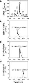
Figure 2. UPLC-MS analysis of the structure of 6
(A) shows UPLC resolution of 6 into six peaks (p1 to p6), which arise from variable sulfation of the PGG scaffold. (B) – (D) show SIR monitoring of 6 at 1388, 1479 and 1569 m/z to identify the peaks corresponding to 9, 10 and 11 sulfated PGG species. Similar SIR profiles were measured for 7, 8 and 12 sulfated species (see Figure S6). See text for detailed interpretation.
Figure 3
Figure 3. Direct inhibition of coagulation and digestive proteases by 6
The inhibition of factor XIa (■), factor Xa (□), thrombin (△), factor XIIa (●), factor IXa (○), factor VIIa (▲), chymotrypsin (◊), and trypsin (♦) by 6 was studied as described in “Experimental Procedures”. Solid lines represent sigmoidal dose–response fits (Eq. 1) to the data to obtain the values of IC50, ΔY, and HS.
Figure 4
Figure 4. Time course of FIX activation by FXIa in the presence and absence of 6
FIX (500 nM) in the assay buffer was incubated with FXIa (3 nM) and 6 (0 or 3 ±M) and aliquots of the reaction analyzed using standard denaturing polyacrylamide gel electrophoresis followed by Western blotting. See Experimental Procedures for details.
Figure 5
Figure 5. Michaelis–Menten kinetics of S-2366 hydrolysis by factor XIa in the presence of 6
The initial rate of hydrolysis at various substrate concentrations was measured in pH 7.4 buffer as described in “Experimental Procedures” using FXIa wild type (FXIa-WT) (A) and FXIa catalytic domain (FXIa-CD) (B). The concentrations of 6 chosen in the study were in (A) 0 (♦), 0.005 (□), 0.5 (▲), 2.5 (○), 5 (■), 50 µg/mL (△) and in (B) 0 (♦), 0.005 (□), 0.5 (▲), 5 µg/mL (○). Solid lines represent nonlinear regressional fits into the data by the Michaelis–Menten Eq. 2.
Figure 6
Figure 6. Competitive direct inhibition of factor XIa by 6 in the presence of UFH (A) and comparison of the predicted and experimentally measured IC50 (B)
Shown in A is the inhibition of factor XIa by 6 in the presence of UFH which was determined spectrophotometrically at pH 7.4 and 37 °C. Solid lines represent fits by the dose–response Eq. 1 to obtain the IC50,predicted, as described in “Experimental Procedures”. The concentrations of UFH selected for the study were 0 (▲), 0.13 (□), 3.3 (●), 33.3 µM (◊). Shown in B is comparison of the predicted and experimentally measured IC50 FXIa inhibition by 6 in the presence of UFH. Open bars represent the measured values, whereas closed bars are the values predicted using Dixon-Webb Eq.4.
Figure 7
Figure 7. Effect of 6 on the clotting times of PT and APTT in human plasma
Prolongation of clotting time as a function of 6 concentration in either prothrombin time assay (PT) (○) or activated partial thromboplastin time assay (APTT) (●). Solid lines are trend lines from which the concentration necessary to double clotting time was deduced. Clotting assays were performed as described in “Experimental Procedures”.
Figure 8
Figure 8. Thromboelastography analysis of clot formation in the presence of 6
Comparison of the effect of 6 and enoxaparin on clot formation in whole blood using thromboelastography analysis. A typical thromboelastogram expected of any anticoagulant is described by R, α, MA, and G parameters. This analysis was performed as described in “Experimental Procedures”.

Similar articles

Cited by

References

    1. Henry BL, Desai UR. Anticoagulants: Drug discovery and development. In: Rotella D, Abraham DJ, editors. Burger’s Medicinal Chemistry. 7th ed. New York: John Wiley and Sons; 2010. pp. 365–408.
    1. Bates SM, Weitz JI. The status of new anticoagulants. Br. J. Haematol. 2006;134:3–19. - PubMed
    1. Hirsh J, Anand SS, Halperin JL, Fuster V. Guide to anticoagulant therapy: Heparin: a statement for healthcare professionals from the American Heart Association. Circulation. 2001;103:2994–3018. - PubMed
    1. Ansell J, Hirsh J, Hylek E, Jacobson A, Crowther M, Palareti G. Pharmacology and management of the vitamin K antagonists: American College of Chest Physicians. Evidence-based clinical practice guidelines, 8th ed. Chest. 2008;133:160S–198S. - PubMed
    1. Eikelboom JW, Wallentin L, Connolly SJ, Ezekowitz M, Healey JS, Oldgren J, Yang S, Alings M, Kaatz S, Hohnloser SH, Diener H-C, Franzosi MG, Huber K, Reilly P, Varrone J, Yusuf S. Risk of Bleeding With 2 Doses of Dabigatran Compared With Warfarin in Older and Younger Patients With Atrial Fibrillation. An analysis of the randomized evaluation of long-term anticoagulant therapy (RE-LY) Trial. Circulation. 2011;123:2363–2372. - PubMed

Publication types

MeSH terms