

Physiology of the intrathecal bolus: the leptomeningeal route for macromolecule and particle delivery to CNS
- PMID: 23316936
- PMCID: PMC3646927
- DOI: 10.1021/mp300474m
Physiology of the intrathecal bolus: the leptomeningeal route for macromolecule and particle delivery to CNS
Abstract
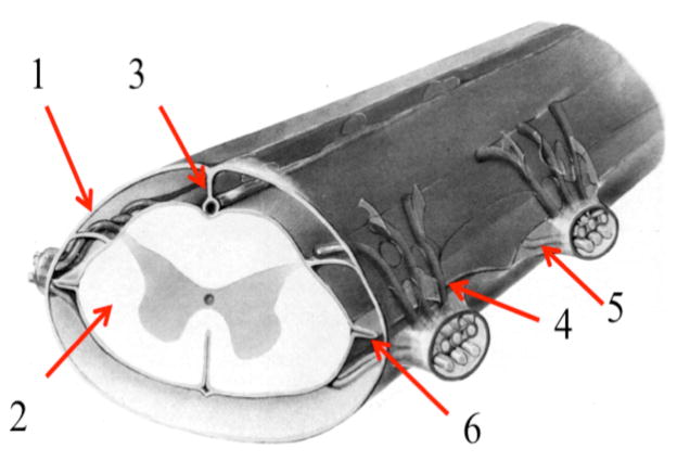
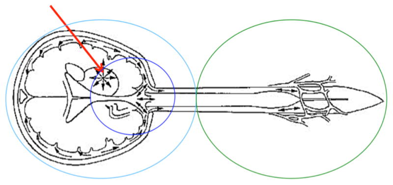
Presently, there are no effective treatments for several diseases involving the CNS, which is protected by the blood-brain, blood-CSF, and blood-arachnoid barriers. Traversing any of these barriers is difficult, especially for macromolecular drugs and particulates. However, there is significant experimental evidence that large molecules can be delivered to the CNS through the cerebrospinal fluid (CSF). The flux of the interstitial fluid in the CNS parenchyma, as well as the macro flux of CSF in the leptomeningeal space, are believed to be generally opposite to the desirable direction of CNS-targeted drug delivery. On the other hand, the available data suggest that the layer of pia mater lining the CNS surface is not continuous, and the continuity of the leptomeningeal space (LMS) with the perivascular spaces penetrating into the parenchyma provides an unexplored avenue for drug transport deep into the brain via CSF. The published data generally do not support the view that macromolecule transport from the LMS to CNS is hindered by the interstitial and CSF fluxes. The data strongly suggest that leptomeningeal transport depends on the location and volume of the administered bolus and consists of four processes: (i) pulsation-assisted convectional transport of the solutes with CSF, (ii) active "pumping" of CSF into the periarterial spaces, (iii) solute transport from the latter to and within the parenchyma, and (iv) neuronal uptake and axonal transport. The final outcome will depend on the drug molecule behavior in each of these processes, which have not been studied systematically. The data available to date suggest that many macromolecules and nanoparticles can be delivered to CNS in biologically significant amounts (>1% of the administered dose); mechanistic investigation of macromolecule and particle behavior in CSF may result in a significantly more efficient leptomeningeal drug delivery than previously thought.
Figures


References
Publication types
MeSH terms
Substances
Grants and funding
LinkOut - more resources
Full Text Sources
Other Literature Sources
